Review
. 2023 Feb;39(1):111-143.
doi: 10.1007/s10565-022-09773-7. Epub 2022 Sep 16.

Oxygen toxicity: cellular mechanisms in normobaric hyperoxia

Affiliations
Review

Oxygen toxicity: cellular mechanisms in normobaric hyperoxia

Ricardo Alva et al. Cell Biol Toxicol. 2023 Feb.

Abstract

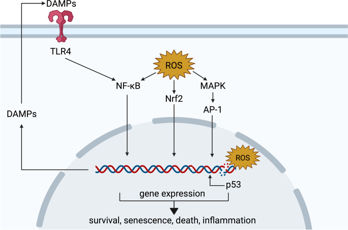
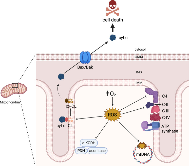
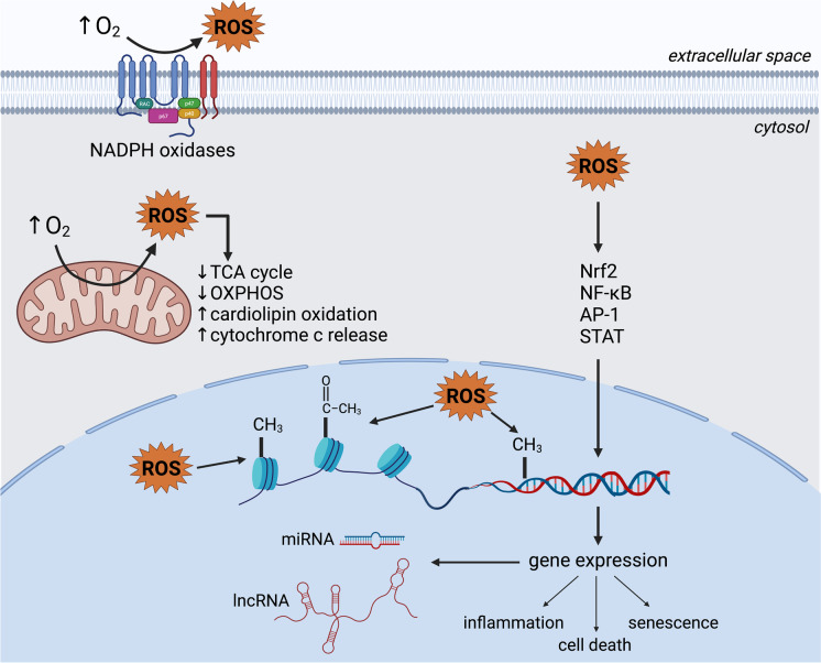
In clinical settings, oxygen therapy is administered to preterm neonates and to adults with acute and chronic conditions such as COVID-19, pulmonary fibrosis, sepsis, cardiac arrest, carbon monoxide poisoning, and acute heart failure. In non-clinical settings, divers and astronauts may also receive supplemental oxygen. In addition, under current standard cell culture practices, cells are maintained in atmospheric oxygen, which is several times higher than what most cells experience in vivo. In all the above scenarios, the elevated oxygen levels (hyperoxia) can lead to increased production of reactive oxygen species from mitochondria, NADPH oxidases, and other sources. This can cause cell dysfunction or death. Acute hyperoxia injury impairs various cellular functions, manifesting ultimately as physiological deficits. Chronic hyperoxia, particularly in the neonate, can disrupt development, leading to permanent deficiencies. In this review, we discuss the cellular activities and pathways affected by hyperoxia, as well as strategies that have been developed to ameliorate injury. • Hyperoxia promotes overproduction of reactive oxygen species (ROS). • Hyperoxia dysregulates a variety of signaling pathways, such as the Nrf2, NF-κB and MAPK pathways. • Hyperoxia causes cell death by multiple pathways. • Antioxidants, particularly, mitochondria-targeted antioxidants, have shown promising results as therapeutic agents against oxygen toxicity in animal models.

Keywords: Antioxidants; Cell death; Hyperoxia; Mitochondria; Oxygen toxicity; Reactive oxygen species.


Conflict of interest statement

The authors declare no competing interests.

Figures

Fig. 1
Fig. 1
Signaling pathways affected in hyperoxia. Excessive ROS modulate intracellular signaling, including via nuclear factor erythroid 2-related factor 2 (Nrf2), nuclear factor kappa-light-chain-enhancer of activated B cells (NF-κB), and mitogen-activated protein kinase (MAPK) pathways. In parallel, oxidative damage to DNA activates p53, which induces the transcription of target genes. In turn, damage-associated molecular patterns (DAMPs) upregulated by these pathways are released into the extracellular space, where they can bind receptors such as the toll-like receptor 4 (TLR4) and further activate the NF-κB pathway. Signaling events orchestrated by these and other pathways determine the outcome of hyperoxia-mediated oxidative stress and may include cell survival, senescence, death, and inflammation. Created with BioRender.com
Fig. 2
Fig. 2
Mitochondrial targets of hyperoxia-mediated injury. Hyperoxia drives the over production of mitochondrial reactive oxygen species (ROS), which inhibit metabolic enzymes such as aconitase, α-ketoglutarate dehydrogenase (α-KGDH), and pyruvate dehydrogenase (PDH), and respiratory complexes I and II, leading to bioenergetic failure. mROS oxidize mitochondrial DNA (mtDNA) and cardiolipin (CL), further promoting dysfunction and leading to the release of cytochrome c (cyt c) into the cytosol through Bcl-2-associated X protein/Bcl-2 homologous antagonist killer (Bax/Bak) oligomers to instigate apoptosis. Created with BioRender.com
Fig. 3
Fig. 3
Molecular mechanisms and cellular pathways of hyperoxia. Through an increased production of reactive oxygen species, hyperoxia dysregulates signaling pathways and promotes epigenetic modifications, resulting in altered gene expression, and ultimately leading to senescence, inflammation, and death. In the mitochondria, hyperoxia inhibits respiration and promotes cardiolipin oxidation and cytochrome c release, further contributing to the induction of cell death pathways. Created with BioRender.com

References

    1. Abbas M, Moradi F, Hu W, Regudo KL, Osborne M, Pettipas J, et al. Vertebrate cell culture as an experimental approach – limitations and solutions. Comp Biochem Physiol B Biochem Mol Biol. 2021;254:110570. doi: 10.1016/j.cbpb.2021.110570. - DOI - PubMed
    1. Acehan D, Malhotra A, Xu Y, Ren M, Stokes DL, Schlame M. Cardiolipin affects the supramolecular organization of ATP synthase in mitochondria. Biophys J. 2011;100:2184–2192. doi: 10.1016/j.bpj.2011.03.031. - DOI - PMC - PubMed
    1. Agarwal S, Sohal RS. DNA oxidative damage and life expectancy in houseflies. Proc Natl Acad Sci U S A. 1994;91:12332–12335. doi: 10.1073/pnas.91.25.12332. - DOI - PMC - PubMed
    1. Aguilera O, Fernández AF, Muñoz A, Fraga MF. epigenetics and environment: a complex relationship. J Appl Physiol. 2010;109:243–251. doi: 10.1152/japplphysiol.00068.2010. - DOI - PubMed
    1. Alam MA, Betal SGnee, Aghai ZH, Bhandari V. Hyperoxia causes miR199a-5p-mediated injury in the developing lung. Pediatr Res. 2019;86:579–588. doi: 10.1038/s41390-019-0524-3. - DOI - PubMed

Publication types