. 2022 Dec;11(6):1951-1959.
doi: 10.1007/s40123-022-00570-3. Epub 2022 Sep 16.

Pole to Pole Surgery in Ocular Trauma: Standardizing Surgical Steps

Affiliations

Pole to Pole Surgery in Ocular Trauma: Standardizing Surgical Steps

Rino Frisina et al. Ophthalmol Ther. 2022 Dec.

Abstract

This commentary describes steps in ocular reconstruction surgery following ocular globe injuries in both the anterior and posterior segment causing corneal opacity and aphakia. The authors propose to reorder the sequence of surgical manoeuvres during pars plana vitrectomy combined with keratoplasty and aphakia treatment without capsular support and highlight the advantages in the choice of the intraocular lens to implant. A mental outline of all surgical manoeuvres, being aware of the complications that can arise during surgery and knowing the long-term benefits of making more careful choices, can make this surgery more effective and safer.

Keywords: Aphakia; Keratoplasty; Ocular globe injuries; Pars plana vitrectomy.


Figures

Fig. 1
Fig. 1
In the figure the author reported a case of open globe trauma with foreign body; the patient underwent pars plana vitrectomy (PPV) combined with penetrating keratoplasty (PK) according to the proposed surgical technique. a Magnetic resonance (MRI) image of a patient affected by open globe trauma with foreign body. The image shows the measurement of the length and thickness of the foreign body, a glass piece. b Eye before and after removal of corneal sutures. The corneal topography was used to evaluate the corneal morphological changes after removal of corneal sutures; c shows the astigmatism after PK and the reduction of the astigmatism after removal of corneal sutures. d Pupillometry study in scotopic and mesopic conditions showing a permanent traumatic mydriasis. r Anterior chamber depth (ACD) measurement. In the reported case the ACD was superior to 3 mm with a wide-open anterior chamber angle (f) thanks to the position of the IOL in the posterior chamber, fixated to the sclera. The authors monitored endothelial cell count (g) with an early reduction and a stabilization around 1000 cells/mm2 after 24 postoperative months
Fig. 2
Fig. 2
Temporal sequence of the surgical steps from the trocar insertion (a, b), to the preparation of the scleral pocket (c) and finally to the corneal trephination and keratoprosthesis placement (df)
Fig. 3
Fig. 3
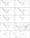
Temporal sequence of the surgical steps of the scleral fixation technique to implant the intraocular lens (ak)

References

    1. Yucel OE, Demir S, Niyaz L, Sayin O, Gul A, Ariturk N. Clinical characteristics and prognostic factors of scleral rupture due to blunt ocular trauma. Eye (Lond) 2016;12:1606–1613. doi: 10.1038/eye.2016.194. - DOI - PMC - PubMed
    1. Négrel AD, Thylefors B. The global impact of eye injuries. Ophthalmic Epidemiol. 1998;3:143–169. doi: 10.1076/opep.5.3.143.8364. - DOI - PubMed
    1. Kenyon KR, Starck T, Hersh PS. Penetrating keratoplasty and for severe ocular trauma. Ophthalmology. 1992;99(3):396–402. doi: 10.1016/s0161-6420(92)31961-x. - DOI - PubMed
    1. Petrelli M, Schmutz L, Gkaragkani E, Droutsas K, Kymionis GD. Simultaneous penetrating keratoplasty and implantation of a new scleral-fixated, sutureless, posterior chamber intraocular lens (Soleko, Carlevale): a novel technique. Cornea. 2020;39(11):1450–1452. doi: 10.1097/ICO.0000000000002378. - DOI - PubMed
    1. Al-qahtani FA. Scleral fixation of intraocular lenses combined with penetrating keratoplasty. J Cataract Refract Surg. 2010;36(3):373–376. doi: 10.1016/j.jcrs.2009.09.041. - DOI - PubMed

LinkOut - more resources