. 2022 Nov 7;219(11):e20220514.
doi: 10.1084/jem.20220514. Epub 2022 Sep 16.

Autoantibodies against type I IFNs in patients with critical influenza pneumonia

Qian Zhang #  1   2   3 Andrés Pizzorno #  4 Lisa Miorin #  5   6 Paul Bastard #  1   2   3   7 Adrian Gervais #  2   3 Tom Le Voyer #  2   3 Lucy Bizien #  2   3 Jeremy Manry  2   3 Jérémie Rosain  2   3 Quentin Philippot  2   3 Kelian Goavec  2   3 Blandine Padey  4   8 Anastasija Cupic  5 Emilie Laurent  4   9 Kahina Saker  10 Martti Vanker  11 Karita Särekannu  11 COVID Human Genetic EffortEtablissement Français du Sang Study GroupConstances Cohort3C-Dijon StudyCerba HealthCare GroupLyon Antigrippe Working GroupREIPI INF Working GroupTamara García-Salum  12   13 Marcela Ferres  12 Nicole Le Corre  12 Javier Sánchez-Céspedes  14   15   16 María Balsera-Manzanero  14   15   16 Jordi Carratala  14   17   18 Pilar Retamar-Gentil  14   16   19 Gabriela Abelenda-Alonso  17   20 Adoración Valiente  14   15   19 Pierre Tiberghien  21 Marie Zins  22 Stéphanie Debette  23 Isabelle Meyts  24 Filomeen Haerynck  25 Riccardo Castagnoli  26 Luigi D Notarangelo  26 Luis I Gonzalez-Granado  27 Nerea Dominguez-Pinilla  28 Evangelos Andreakos  29 Vasiliki Triantafyllia  29 Carlos Rodríguez-Gallego  30   31 Jordi Solé-Violán  31   32   33 José Juan Ruiz-Hernandez  34 Felipe Rodríguez de Castro  35   36 José Ferreres  37   38 Marisa Briones  39 Joost Wauters  40 Lore Vanderbeke  40 Simon Feys  40 Chen-Yen Kuo  41   42 Wei-Te Lei  41   43 Cheng-Lung Ku  41   44   45 Galit Tal  46   47 Amos Etzioni  46 Suhair Hanna  46 Thomas Fournet  48 Jean-Sebastien Casalegno  49 Gregory Queromes  4 Laurent Argaud  50 Etienne Javouhey  51 Manuel Rosa-Calatrava  4   9 Elisa Cordero  14   15   16   52 Teresa Aydillo  5   6 Rafael A Medina  5   12 Kai Kisand #  11 Anne Puel #  1   2   3 Emmanuelle Jouanguy #  1   2   3 Laurent Abel #  1   2   3 Aurélie Cobat #  1   2   3 Sophie Trouillet-Assant #  4   10 Adolfo García-Sastre #  5   6   53   54   55 Jean-Laurent Casanova #  1   2   3   7   56
Collaborators, Affiliations

Autoantibodies against type I IFNs in patients with critical influenza pneumonia

Qian Zhang et al. J Exp Med. .

Abstract

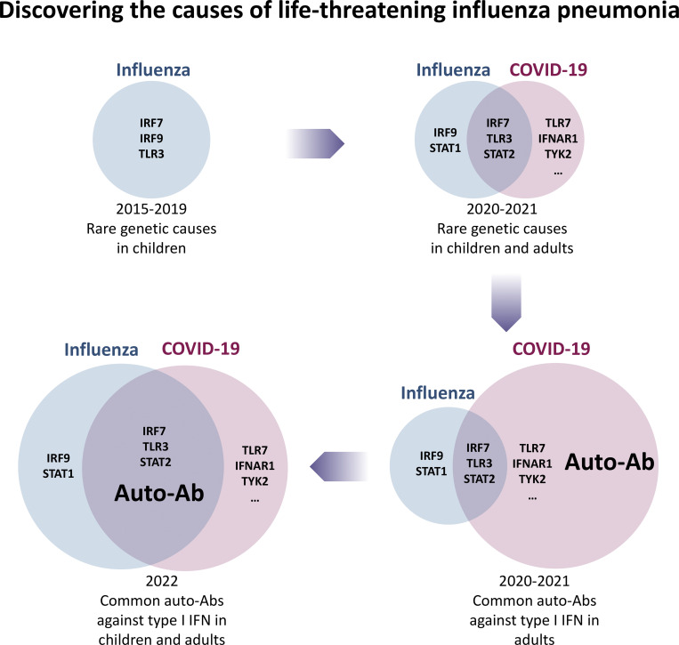
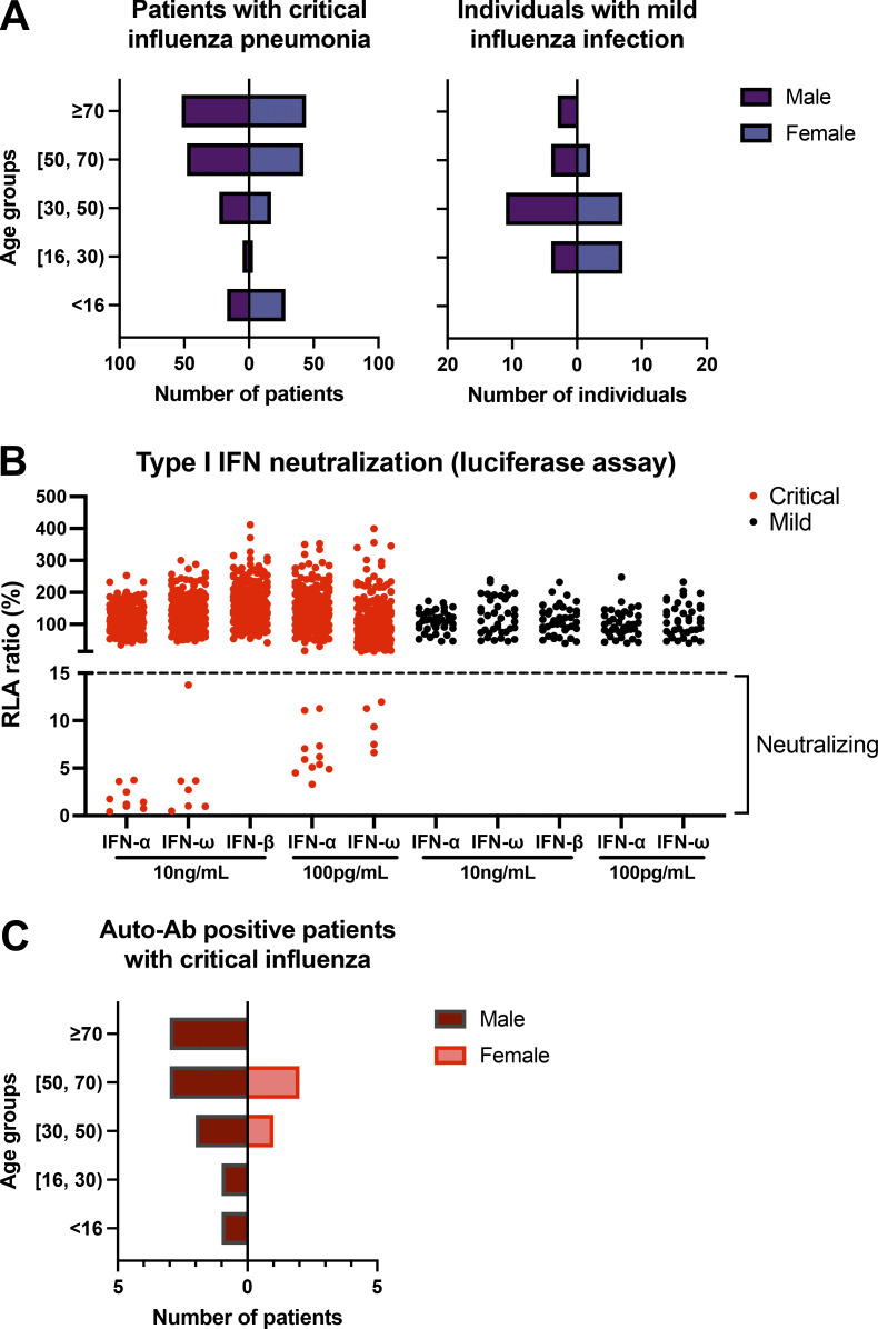
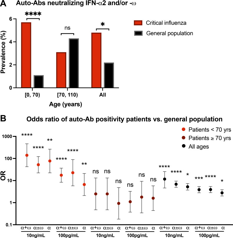
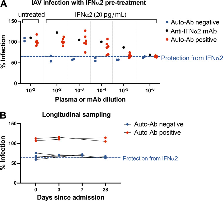
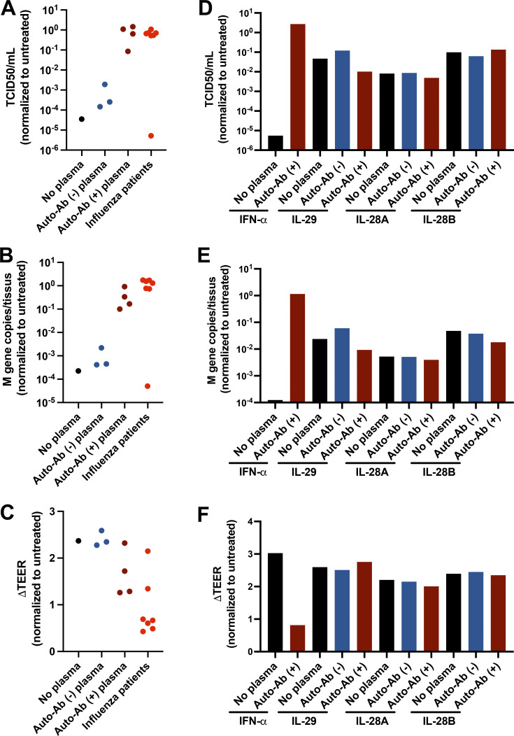
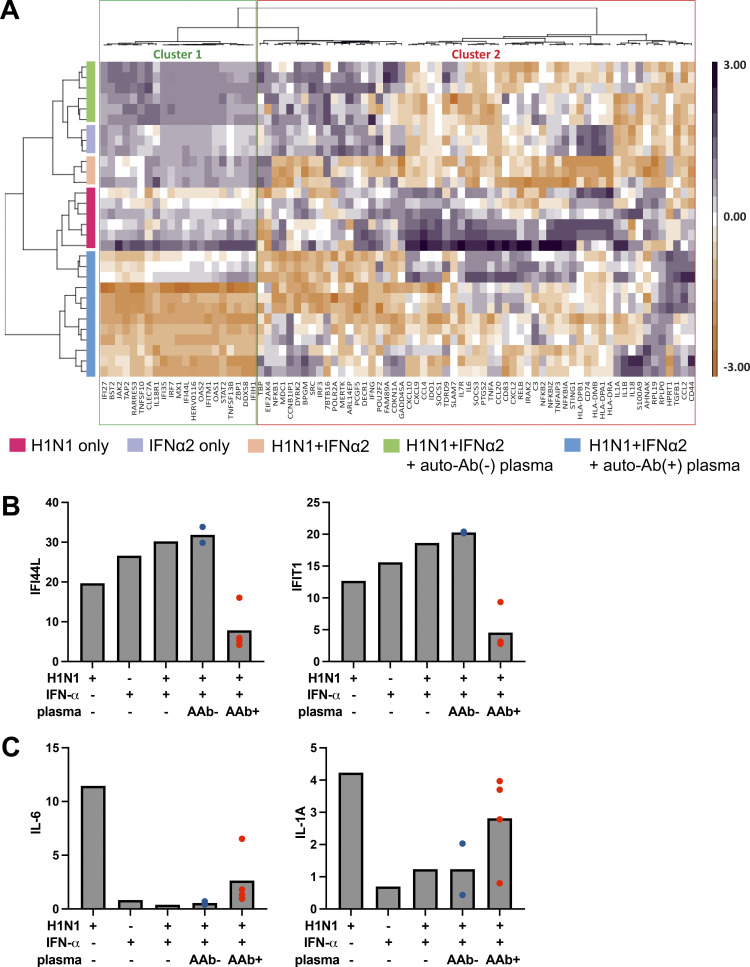
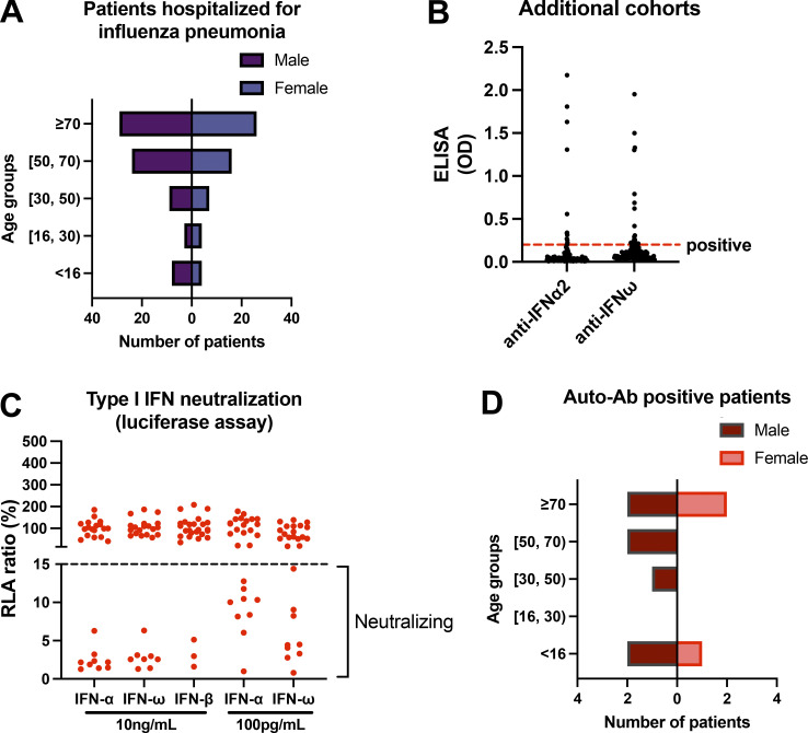
Autoantibodies neutralizing type I interferons (IFNs) can underlie critical COVID-19 pneumonia and yellow fever vaccine disease. We report here on 13 patients harboring autoantibodies neutralizing IFN-α2 alone (five patients) or with IFN-ω (eight patients) from a cohort of 279 patients (4.7%) aged 6-73 yr with critical influenza pneumonia. Nine and four patients had antibodies neutralizing high and low concentrations, respectively, of IFN-α2, and six and two patients had antibodies neutralizing high and low concentrations, respectively, of IFN-ω. The patients' autoantibodies increased influenza A virus replication in both A549 cells and reconstituted human airway epithelia. The prevalence of these antibodies was significantly higher than that in the general population for patients <70 yr of age (5.7 vs. 1.1%, P = 2.2 × 10⁻⁵), but not >70 yr of age (3.1 vs. 4.4%, P = 0.68). The risk of critical influenza was highest in patients with antibodies neutralizing high concentrations of both IFN-α2 and IFN-ω (OR = 11.7, P = 1.3 × 10⁻⁵), especially those <70 yr old (OR = 139.9, P = 3.1 × 10⁻¹⁰). We also identified 10 patients in additional influenza patient cohorts. Autoantibodies neutralizing type I IFNs account for ∼5% of cases of life-threatening influenza pneumonia in patients <70 yr old.


Conflict of interest statement

Disclosures: A. Pizzorno reported a patent to WO2016/146836 licensed (Signia Therapeutics), a patent to WO2017/174593 licensed (Signia Therapeutics), and a patent to WO2019/224489 licensed (Signia Therapeutics); and is the co-founder of Signia Therapeutics SAS. N. Le Corre reported personal fees from SINOVAC outside the submitted work. P. Retamar-Gentil reported personal fees from Merck outside the submitted work. I. Meyts reported grants from CSL-Behring outside the submitted work. E. Andreakos reported grants from Janssen Pharmaceuticals during the conduct of the study. J. Wauters reported grants and personal fees from Pfizer and Gilead outside the submitted work. L. Vanderbeke reported grants from Research Foundation Flanders and non-financial support from Pfizer outside the submitted work. S. Feys reported grants from Pfizer outside the submitted work. J. Casalegno reported “other” from Pfizer and grants from Sanofi outside the submitted work. M. Rosa-Calatrava reported a patent to WO2016/146836 licensed (Signia Therapeutics), a patent to WO2017/174593 licensed (Signia Therapeutics), and a patent to WO2019/224489 licensed (Signia Therapeutics); and is the co-founder of Signia Therapeutics SAS. S. Trouillet-Assant reported non-financial support from BioMérieux outside the submitted work. A. Garcia-Sastre reported “other” from Vivaldi Biosciences, Pagoda, Contrafect, Vaxalto, Accurius, Curelab oncology, and Curelab veterinary; personal fees from Avimex, 7Hills, Esperovax, Pfizer, Farmak, Applied Biological Laboratories, Paratus, Pharmamar, Pfizer, and Synairgen; grants from Pfizer, Pharmamar, Blade Therapeutics, Avimex, Accurius, Dynavax, Kenall Manufacturing, ImmunityBio, Nanocomposix, Merck, Model Medicines, Atea Pharma, Shenwa Biosciences, Johnson & Johnson, 7 Hills, Hexamer, N-fold LLC, and Applied Biological Laboratories outside the submitted work; in addition, A. Garcia-Sastre had a patent for influenza virus vaccines and uses thereof issued; and invited speaker in meeting events organized by Seqirus, Janssen, Abbott, and Astrazeneca. J. Casanova reported a patent to PCT/US2021/042741 pending. No other disclosures were reported.

Figures

None
Graphical abstract
Figure 1.
Figure 1.
Auto-Abs neutralizing IFN-α2 and/or IFN-ω in patients with critical influenza pneumonia. (A) Age and sex distribution of the patients with critical influenza pneumonia or mild influenza infection. (B) Luciferase-based neutralization assay to detect auto-Abs neutralizing 10 ng/ml or 100 pg/ml IFN-α2, IFN-ω, or IFN-β. Plasma samples from patients with critical (red) or mild (black) influenza were diluted 1:10 in all tests. HEK293T cells were transfected with the dual luciferase system with IFN-sensitive response elements (ISRE) before treatment with type I IFNs with or without patient plasma, and relative luciferase activity (RLA) was calculated by normalizing firefly luciferase activity against Renilla luciferase activity. An RLA <15% of the value for the mock treatment was considered to correspond to neutralizing activity (dashed line; Bastard et al., 2021a). Experiments were repeated at least twice, and the average was plotted in the figure. (C) Age and sex distribution of patients with auto-Ab neutralizing IFN-α2 and/or IFN-ω (n = 13).
Figure 2.
Figure 2.
Enrichment in auto-Ab–positive cases among patients with critical influenza pneumonia. (A) Prevalence of auto-Ab–positive cases among patients with critical influenza (n = 279, red bars) and in the general population (n = 34,159, black bars). *, P < 0.05; ****, P < 10−5. (B) OR for the presence of auto-Abs, by sex and age, relative to the general population, with adjustment of the comparison by means of Firth’s bias-corrected logistic regression. The horizontal bars indicate the upper and lower limits of the 95% CIs. α + ω, auto-Abs neutralizing both IFN-α2 and IFN-ω; α ± ω, auto-Abs neutralizing IFN-α2 with or without IFN-ω; α, auto-Abs neutralizing IFN-α2 only; *, P < 0.05; **, P < 10−2; ***, P < 10−3; ****, P < 10−4.
Figure 3.
Figure 3.
Neutralizing auto-Abs block the antiviral function of IFN-α2 in IAV-infected A549 epithelial cells. (A) A549 cells were treated with 20 pg/ml exogenous IFN-α2 with or without patient plasma (titrated to the dilutions indicated on the x axis), anti–IFN-α2 monoclonal antibody, and healthy donor plasma overnight before infection with IAV Cal/09 virus expressing NS1-mCherry (CalNSmCherry) at an MOI of 0.5. The day after infection, the percentage of the cells infected was determined with a Celigo (Nexcelcom) imaging cytometer. The dotted line at 64.98% represents the mean percentage infection in cells treated with 20 pg/ml IFN-α2 in the absence of plasma or anti–IFN-α2 antibody. Experiments were repeated four times. (B) Longitudinal testing of six patients with life-threatening influenza pneumonia (two positive and four negative for auto-Abs), with the assay as described in A.
Figure 4.
Figure 4.
Neutralizing auto-Abs block the antiviral function of IFN-α2 in IAV-infected HAE cultures. (A–F) HAE reconstituted from human nasal primary cells and maintained in an air–liquid interface were either left untreated or treated with 2 ng/ml exogenous IFN-α2a (A–C) or 20 ng/ml exogenous IL-29, IL-28A, or IL-28B (D–F), in the presence of inactivated patient plasma (1:100 diluted) for 24 h before IAV infection. Cells were treated again on the basolateral side with same concentration of IFN-α2a or IFN-λ1/IL-29, IFN-λ2/IL-28A, or IFN-λ3/IL-28B in the presence of patient plasma (n = 7) 1 h after IAV infection. These seven patients had auto-Abs neutralizing IFN-α at 10 ng/ml, but not IFN-β or -λ. HAE apical poles were washed, 54 h after infection, and titrated by TCID50 determination (A and D) and quantitative RT-PCR (B and E). Changes in TEER (ΔTEER) were measured as a surrogate for the integrity of HAE (C and F). Previously identified auto-Ab–positive (auto-Ab [+]) or auto-Ab [−] plasma samples were used as controls. Experiments were repeated three times.
Figure 5.
Figure 5.
ISG and proinflammatory responses in IAV-infected HAE cultures. HAE reconstituted from human nasal primary cells and maintained in an air–liquid interface were either left untreated or treated with 2 ng/ml exogenous IFN-α2a in the presence of inactivated patient plasma (1:100 dilution) for 24 h before IAV infection. Cells were treated again on the basolateral side with 2 ng/ml IFN-α2a in the presence of patient plasma 1 h after IAV infection. RNA was isolated 54 h after infection, and NanoString analysis was performed with a panel of immune response genes. (A) Heatmap of gene expression profiles from unsupervised analysis (Euclidean distance matrix, Ward’s method) generated by scaling and centering log10-transformed normalized gene expression (expressed as fold-change induction relative to mock conditions) and based on the full 96-gene panel. Gene and sample clustering is indicated by dendrogram trees above and to the left, respectively, of the heatmap. Gene clustering distinguished ISGs (cluster 1) from proinflammatory genes (cluster 2; Table S1). (B and C) Relative expression (mean) levels of two ISGs, IFI44L and IFIT1 (B), and two proinflammatory cytokines, IL-6 and IL1A (C), based on NanoString analysis on total cellular RNA extracted after infection. Gene expression is expressed as a fold-change induction relative to mock conditions (untreated/uninfected). AAb−, auto-Ab–negative plasma; AAb+, auto-Ab–positive plasma.
Figure 6.
Figure 6.
Auto-Abs neutralizing IFN-α2 and/or IFN-ω in ELISA-positive patients hospitalized with influenza pneumonia in additional cohorts. (A) Age and sex distribution of patients from Chile, Spain, France, Belgium, and Taiwan hospitalized for influenza pneumonia (n = 130). (B) Patient plasma samples were tested by ELISA for auto-Abs against IFN-α2 and -ω. Patient plasma samples were diluted 1:50 before being added to plates coated with 2 μg/ml rhIFN-α or rhIFN-ω. HRP-conjugated goat antiserum against human IgG or IgA was added to final concentration of 2 μg/ml. OD was measured. Each plasma sample was tested once. (C) Luciferase-based neutralization assay to detect auto-Abs neutralizing 10 ng/ml or 100 pg/ml IFN-α2, IFN-ω, or IFN-β. Plasma samples from ELISA-positive patients were diluted 1:10 in all tests. HEK293T cells were transfected with the dual luciferase system with IFN-sensitive response elements (ISRE) before treatment with type I IFNs with or without plasma from patients, and relative luciferase activity (RLA) was calculated by normalizing firefly luciferase activity against Renilla luciferase activity. An RLA <15% the value for the mock treatment was considered to indicate that the antibodies were neutralizing (dashed line). (D) Age and sex distribution of patients with auto-Ab neutralizing IFN-α2 and/or IFN-ω (n = 10).

References

    1. Abers, M.S., Rosen L.B., Delmonte O.M., Shaw E., Bastard P., Imberti L., Quaresima V., Biondi A., Bonfanti P., Castagnoli R., et al. . 2021. Neutralizing type-I interferon autoantibodies are associated with delayed viral clearance and intensive care unit admission in patients with COVID-19. Immunol. Cell Biol. 99:917–921. 10.1111/imcb.12495 - DOI - PMC - PubMed
    1. Abolhassani, H., Landegren N., Bastard P., Materna M., Modaresi M., Du L., Aranda-Guillen M., Sardh F., Zuo F., Zhang P., et al. . 2022. Inherited IFNAR1 deficiency in a child with both critical COVID-19 pneumonia and multisystem inflammatory syndrome. J. Clin. Immunol. 42:471–48310.1007/s10875-022-01215-7 - DOI - PMC - PubMed
    1. Acosta-Ampudia, Y., Monsalve D.M., Rojas M., Rodriguez Y., Gallo J.E., Salazar-Uribe J.C., Santander M.J., Cala M.P., Zapata W., Zapata M.I., et al. . 2021. COVID-19 convalescent plasma composition and immunological effects in severe patients. J. Autoimmu. 118:102598. 10.1016/j.jaut.2021.102598 - DOI - PMC - PubMed
    1. Akbil, B., Meyer T., Stubbemann P., Thibeault C., Staudacher O., Niemeyer D., Jansen J., Mühlemann B., Doehn J., Tabeling C., et al. ; Pa-COVID study Group . 2022. Early and Rapid Identification of COVID-19 Patients with Neutralizing Type I Interferon Auto-antibodies. J. Clin. Immunol. 10.1007/s10875-022-01252-2 - DOI - PMC - PubMed
    1. Asano, T., Boisson B., Onodi F., Matuozzo D., Moncada-Velez M., Maglorius Renkilaraj M.R.L., Zhang P., Meertens L., Bolze A., Materna M., et al. . 2021. X-linked recessive TLR7 deficiency in ∼1% of men under 60 years old with life-threatening COVID-19. Sci. Immunol. 6:eabl4348. 10.1126/sciimmunol.abl4348 - DOI - PMC - PubMed

Publication types