. 2022 Sep 16;8(37):eabh3260.
doi: 10.1126/sciadv.abh3260. Epub 2022 Sep 16.

Activation of TrkB-Akt signaling rescues deficits in a mouse model of SCA6

Affiliations

Activation of TrkB-Akt signaling rescues deficits in a mouse model of SCA6

Anna A Cook et al. Sci Adv. .

Abstract

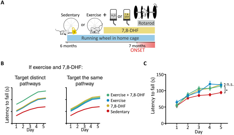
Spinocerebellar ataxia type 6 (SCA6) is a neurodegenerative disease resulting in motor coordination deficits and cerebellar pathology. Expression of brain-derived neurotrophic factor (BDNF) is reduced in postmortem tissue from SCA6 patients. Here, we show that levels of cerebellar BDNF and its receptor, tropomyosin receptor kinase B (TrkB), are reduced at an early disease stage in a mouse model of SCA6 (SCA684Q/84Q). One month of exercise elevated cerebellar BDNF expression and improved ataxia and cerebellar Purkinje cell firing rate deficits. A TrkB agonist, 7,8-dihydroxyflavone (7,8-DHF), likewise improved motor coordination and Purkinje cell firing rate and elevated downstream Akt signaling. Prolonged 7,8-DHF administration persistently improved ataxia when treatment commenced near disease onset but was ineffective when treatment was started late. These data suggest that 7,8-DHF, which is orally bioavailable and crosses the blood-brain barrier, is a promising therapeutic for SCA6 and argue for the importance of early intervention for SCA6.


Figures

Fig. 1.
Fig. 1.. SCA684Q/84Q mice have reduced cerebellar BDNF and TrkB at disease onset.
(A) Schematic of sample collection for BDNF ELISA. (B) At 5 to 6 months, the level of BDNF (pg/mg of soluble protein) in the cerebellar vermis of SCA684Q/84Q mice is not significantly different from WT. However, at 7 months (C) and 12 months (D), there is a significant reduction in BDNF levels detected with an ELISA in cerebellar vermis tissue in SCA684Q/84Q mice compared to WT (BDNF pg/mg soluble protein). (E) Representative images show that BDNF is expressed in the cerebellar cortex at 5 to 6 months but is decreased in SCA684Q/84Q mice. Scale bar, 50 μm. (F) Quantification shows that BDNF levels are reduced in the Purkinje cell somata, the molecular layer, and the granule cell layer in SCA684Q/84Q mice. (G) Representative images show that BDNF is expressed in the cerebellar cortex at 7 months, but expression is decreased in SCA684Q/84Q mice. Scale bar, 50 μm. (H) Quantification showing that BDNF levels are reduced in all the three cortical layers: Purkinje cell layer, the molecular layer, and the granule cell layer. (I) Representative images show that TrkB is expressed in the cerebellar cortex at 7 months, but expression is decreased in SCA684Q/84Q mice. Scale bar, 50 μm. (J) Quantification of TrkB expression in the three cortical layers. TrkB is reduced in the Purkinje cell layer but not in the molecular layer or the granule cell layer. All statistical tests and P values are reported in table S2. ***P < 0.005; **P < 0.01; *P < 0.05; not significant (n.s.), P > 0.05. All data points are normalized to the median WT value of the group.
Fig. 2.
Fig. 2.. Exercise increases cerebellar BDNF levels in SCA684Q/84Q mice.
(A) Schematic of exercise protocol. IHC, immunohistochemistry. (B) BDNF immunoreactivity in sedentary (left) and exercise (right) WT (top) and SCA684Q/84Q (SCA6, bottom) mice at 7 months. Scale bars, 50 μm. (C to E) Cerebellar BDNF expression in cortical layers: (C) Purkinje cell somata, (D) the molecular layer, and (E) the granule cell layer. (F) Cerebellar TrkB expression in sedentary (left) and exercised (right) WT (top) and SCA684Q/84Q (bottom) mice at 7 months. Scale bars, 100 μm. (G to I) Quantification of changes in TrkB expression. (G) Purkinje cell, (H) molecular layer, and (I) the granule cell layer. All statistical tests and P values are reported in table S2. *P < 0.05, **P < 0.01, ***P < 0.005, P > 0.05 when comparison is not shown. All data points are normalized to the median WT value of the group.
Fig. 3.
Fig. 3.. Exercise rescues motor coordination and Purkinje cell firing deficits in SCA684Q/84Q mice.
(A) Schematic of exercise protocol. (B) Motor coordination is assessed with an accelerating rotarod assay after 1 month of voluntary wheel running by measuring the latency to fall for 5 days of testing. Latency to fall is significantly different between WT and SCA684Q/84Q mice, SCA684Q/84Q and SCA684Q/84Q exercise mice, and SCA684Q/84Q exercise and WT mice. (C) Sample juxtacellular Purkinje cell recordings from WT (left) and SCA684Q/84Q (right) sedentary (top) or exercise (bottom) mouse cerebellar acute slices. (D) Purkinje cell firing frequency (left) is restored in SCA684Q/84Q exercise mice. However, action potential regularity (as measured by the CV of interspike intervals) is not restored by exercise. All statistical tests and P values are reported in table S2. *P < 0.05, **P < 0.01, ***P < 0.005, P > 0.05 when not shown.
Fig. 4.
Fig. 4.. The TrkB agonist 7,8-DHF restores ataxia and Purkinje cell firing deficits in SCA684Q/84Q mice at disease onset.
(A) Proposed model of altered BDNF-TrkB signaling in SCA684Q/84Q mice and its subsequent restoration with 7,8-DHF, a TrkB agonist. (B) Schematic of 7,8-DHF administration protocol. (C) 7,8-DHF improves motor coordination in SCA684Q/84Q mice as measured with rotarod assay. (D) Example traces from juxtacellular recordings of Purkinje cell firing at 7 months after 1 month of 7,8-DHF administration. (E) Purkinje cell firing frequency is elevated after 7,8-DHF administration. However, Purkinje cell firing regularity is unchanged with 7,8-DHF administration (CV). All statistical tests and P values are reported in table S2. ***P < 0.005; n.s., P > 0.05.
Fig. 5.
Fig. 5.. Combining exercise with administration of a TrkB agonist provides no further rescue of ataxia in SCA684Q/84Q mice.
(A) Schematic of exercise and 7,8-DHF combination therapy on SCA684Q/84Q mice (SCA6). (B) Schematics of possible outcomes if exercise and 7,8-DHF are additive, suggesting that they act via distinct pathways (left), or if no further improvement is observed in motor coordination when exercise and 7,8-DHF are co-administered, suggesting that they act on the same signaling pathway (right). (C) No additional improvement is observed when exercise and 7,8-DHF administration are combined, and exercise and 7,8-DHF improve motor coordination, similarly suggesting that they act on the same pathway. All statistical tests and P values are reported in table S2. *P < 0.05; n.s., P > 0.05.
Fig. 6.
Fig. 6.. The TrkB agonist 7,8-DHF activates the Akt signaling pathway in SCA684Q/84Q mice.
(A) Top: Schematic of 7,8-DHF administration followed by tissue collection for Western blots and RPPA. Bottom: TrkB and downstream signaling pathways. (B) Total TrkB levels are significantly increased in the cerebellar vermis of SCA684Q/84Q mice after chronic 7,8-DHF administration, while levels of phosphorylated TrkB are slightly increased by 7,8-DHF (C). (D) Schematic of putative Akt signaling downstream of TrkB activation. (E) Total Akt levels are unchanged in vermis tissue after 7,8-DHF administration in SCA684Q/84Q mice, but phosphorylated Akt levels are increased (F and G), indicating Akt activation. (H) Schematic of putative ERK signaling downstream of TrkB activation. (I to K) 7,8-DHF administration does not lead to activation of the ERK signaling pathway in SCA684Q/84Q mice. (L) Representative images showing pAkt(T308) in the Purkinje cells of the cerebellar vermis. Scale bar, 50 μm. (M) Quantification of pAkt(T308) staining showing that levels are increased in the Purkinje cells and molecular layer of SCA684Q/84Q mice that received 7,8-DHF treatment. All statistical tests and P values are reported in table S2. *P < 0.05, **P < 0.01, ***P < 0.005, P > 0.05 when not shown.
Fig. 7.
Fig. 7.. Chronic administration of 7,8-DHF after disease onset leads to a slow improvement of ataxia in SCA684Q/84Q mice.
(A) Schematic of 7,8-DHF administration protocol. (B) SCA684Q/84Q mice (SCA6) were administered 7,8-DHF for 25 days after 5 days of baseline testing. Improvement in motor coordination was observed after ~2 weeks. (C) Motor performance remained elevated for ~2 weeks after 7,8-DHF withdrawal. All statistical tests and P values are reported in table S2. *P < 0.05, **P < 0.01, ***P < 0.005, P > 0.05 when not shown.
Fig. 8.
Fig. 8.. Early, but not late, 7,8-DHF administration rescues deficits at later stages of disease progression in SCA684Q/84Q mice.
(A) Schematic of long-term chronic 7,8-DHF administration in SCA684Q/84Q mice started near disease onset. (B) Mice treated chronically with 7,8-DHF performed better at 7, 9, and 10 months old. (C) Purkinje cell firing rate (left) was no longer restored after 4 months of drug administration, nor was regularity affected. (D) Schematic of 7,8-DHF administration started in late disease progression. (E) Late-administered 7,8-DHF does not restore firing deficits at 10 months when treatment starts after disease onset at 9 months. (F) Purkinje cell firing rate and regularity (CV) are not significantly different after late administration of 7,8-DHF. All statistical tests and P values are reported in table S2. *P < 0.05; ***P < 0.001; n.s., P > 0.05.

References

    1. Zhuchenko O., Bailey J., Bonnen P., Ashizawa T., Stockton D. W., Amos C., Dobyns W. B., Subramony S. H., Zoghbi H. Y., Lee C. C., Autosomal dominant cerebellar ataxia (SCA6) associated with small polyglutamine expansions in the alpha 1A-voltage-dependent calcium channel. Nat. Genet. 15, 62–69 (1997). - PubMed
    1. Solodkin A., Gomez C. M., Spinocerebellar ataxia type 6. Handb. Clin. Neurol. 103, 461–473 (2012). - PubMed
    1. Watase K., Barrett C. F., Miyazaki T., Ishiguro T., Ishikawa K., Hu Y., Unno T., Sun Y., Kasai S., Watanabe M., Gomez C. M., Mizusawa H., Tsien R. W., Zoghbi H. Y., Spinocerebellar ataxia type 6 knockin mice develop a progressive neuronal dysfunction with age-dependent accumulation of mutant CaV2.1 channels. Proc. Natl. Acad. Sci. U.S.A. 105, 11987–11992 (2008). - PMC - PubMed
    1. Jayabal S., Ljungberg L., Erwes T., Cormier A., Quilez S., El Jaouhari S., Watt A. J., Rapid onset of motor deficits in a mouse model of spinocerebellar ataxia type 6 precedes late cerebellar degeneration. eNeuro 2, ENEURO.0094-15.2015 (2015). - PMC - PubMed
    1. Zuccato C., Cattaneo E., Brain-derived neurotrophic factor in neurodegenerative diseases. Nat. Rev. Neurol. 5, 311–322 (2009). - PubMed