. 2022 Sep 16;8(37):eabn6550.
doi: 10.1126/sciadv.abn6550. Epub 2022 Sep 16.

A flexible electronic strain sensor for the real-time monitoring of tumor regression

Affiliations

A flexible electronic strain sensor for the real-time monitoring of tumor regression

Alex Abramson et al. Sci Adv. .

Abstract

Assessing the efficacy of cancer therapeutics in mouse models is a critical step in treatment development. However, low-resolution measurement tools and small sample sizes make determining drug efficacy in vivo a difficult and time-intensive task. Here, we present a commercially scalable wearable electronic strain sensor that automates the in vivo testing of cancer therapeutics by continuously monitoring the micrometer-scale progression or regression of subcutaneously implanted tumors at the minute time scale. In two in vivo cancer mouse models, our sensor discerned differences in tumor volume dynamics between drug- and vehicle-treated tumors within 5 hours following therapy initiation. These short-term regression measurements were validated through histology, and caliper and bioluminescence measurements taken over weeklong treatment periods demonstrated the correlation with longer-term treatment response. We anticipate that real-time tumor regression datasets could help expedite and automate the process of screening cancer therapies in vivo.


Figures

Fig. 1.
Fig. 1.. Flexible autonomous sensors measuring tumor volume regression.
(A) FAST technology. (B) Light microscopy of cracked gold strain sensor at varying strains. Scale bars, 20 μm. (C) An app recording the resistance change in sensor. (D) FAST contains a printed circuit board (PCB), stretchable strain sensors, and a backpack to hold the sensor on the mouse. (E) Resistance changes in sensors when stretched in 10-μm increments from a prestrain of 50% [individual points from five sensors; line, median; one-way analysis of variance (ANOVA) with Tukey’s multiple comparisons test]. (F) Fold change in resistance of strained sensors (individual curves from 10 sensors; bold line, average). (G) Force required to strain sensors of varying SEBS substrate thicknesses (individual curves from 12 to 13 sensors; line, average ± SD). (H) FAST backpack on mouse held in place using Tegaderm. (I) Fold change in resistance of FAST measuring 3D printed ellipsoids comparable to tumors (individual curves from 10 sensors; bold line, average; ratio paired t test). (J and K) Caliper measured in vivo tumor volumes correlate with (J) FAST resistance outputs and (K) bioluminescence average radiance values [(J) 50 points, three FAST sensors (10 to 20 measurements per sensor) and (K) 50 points, one imaging system; lines, best fit linear regressions ±95% confidence interval]. See fig. S4 for further characterization. ***P < 0.001, ****P < 0.0001. Photo credits: Alex Abramson (C, D, and H) and Facebook Design Resources (C).
Fig. 2.
Fig. 2.. FAST sensor detects a decrease in tumor volume sooner than existing methods in HCC827 mouse models treated orally with erlotinib.
(A to C) FAST reads out tumor volume progression or regression continuously at 5-min intervals in (A) Nu/Nu mice with ~100-mm3 subcutaneous HCC827 human lung cancer tumors receiving no treatment and (B and C) mice with ~200-mm3 tumors receiving erlotinib (50 mg/kg) or vehicle treatments at intervals described in the figure. Individual mouse sensor trend lines are presented as seven-point moving averages. (D) FAST sensor measurements over the entire treatment period. (E and F) Erlotinib- and vehicle-treated mice demonstrate significantly different sensor readouts over (E) the entire treatment period and (F) just 5 hours after treatment administration. (G to I) Caliper and (J to L) luminescence imaging confirm the tumor volume measurements recorded by FAST and demonstrate that wearing the FAST device does not affect the outcomes of the treatment experiments. S+, with FAST sensor; S, no FAST sensor; T+, erlotinib treatment; T, vehicle treatment; data are presented as individual data points or curves; bold, average; unpaired two-tailed Student’s t tests. Scale bar, 5 mm.
Fig. 3.
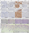
Fig. 3.. Histology from HCC827 tumors treated with erlotinib validates rapid FAST sensor readouts of tumor volume regression.
(A to P) Immunohistochemistry of tumors excised from mice treated for 6 days with vehicle (vehicle), treated for 5 hours with erlotinib, or treated for 6 days with erlotinib. Stains are for cleaved caspase 3 (CC3), a marker associated with cell death; Ki67, a marker associated with cell proliferation; EGFR; and phosphorylated EGFR (pEGFR). Erlotinib is an active inhibitor of EGFR and prevents phosphorylation. (Q to V) Hematoxylin and eosin stains of (Q and R) tumors and (U and V) skin from mice that did or did not wear FAST for 6 days. In the histological sampling, there is no noticeable difference in the cell shapes or distributions between the samples from mice wearing the sensor and the samples from mice not wearing the sensor. Scale bars, 100 μm.
Fig. 4.
Fig. 4.. FAST sensor detects a decrease in tumor volume sooner than existing methods in A20 mouse models treated intratumorally with CpG + anti-OX40.
(A to D) FAST reads out tumor volume progression or regression continuously at 5-min intervals in Balb/c mice with subcutaneous A20 B cell lymphoma tumors receiving 40 μg of CpG and 4 μg of anti-OX40 (n = 5) or vehicle (n = 4) treatments over (A and B) the first few hours following treatment or (C and D) the entire treatment period in the same mice. Individual mouse sensor trend lines are presented as seven-point moving averages. (E and F) Tumor volume measurements using calipers confirm FAST readouts over the entire treatment period. (G to L) Immunohistochemistry of tumors excised from mice treated for 6 days with vehicle (vehicle) or treated once with CpG + anti-OX40 (treated). Staining is against (G and H) CC3, (I and J) Ki67, and (K and L) OX40. The treated stains are from tumors excised within 6 hours after treatment initiation. T+, CpG and anti-OX40 treatment; T, vehicle treatment; S+, with FAST sensor; S, no FAST sensor. Scale bars, 50 μm. Data are presented as individual data point or curves. Bold line, average; (B) unpaired two-tailed Student’s t test; (D and F) one-way ANOVA with Tukey’s multiple comparisons test.

References

    1. Drost J., Clevers H., Organoids in cancer research. Nat. Rev. Cancer 18, 407–418 (2018). - PubMed
    1. Vlachogiannis G., Hedayat S., Vatsiou A., Jamin Y., Fernández-Mateos J., Khan K., Lampis A., Eason K., Huntingford I., Burke R., Rata M., Koh D.-M., Tunariu N., Collins D., Hulkki-Wilson S., Ragulan C., Spiteri I., Moorcraft S. Y., Chau I., Rao S., Watkins D., Fotiadis N., Bali M., Darvish-Damavandi M., Lote H., Eltahir Z., Smyth E. C., Begum R., Clarke P. A., Hahne J. C., Dowsett M., de Bono J., Workman P., Sadanandam A., Fassan M., Sansom O. J., Eccles S., Starling N., Braconi C., Sottoriva A., Robinson S. P., Cunningham D., Valeri N., Patient-derived organoids model treatment response of metastatic gastrointestinal cancers. Science 359, 920–926 (2018). - PMC - PubMed
    1. Clarke M. A., Fisher J., Executable cancer models: Successes and challenges. Nat. Rev. Cancer 20, 343–354 (2020). - PubMed
    1. Lim B., Lin Y., Navin N., Advancing cancer research and medicine with single-cell genomics. Cancer Cell 37, 456–470 (2020). - PMC - PubMed
    1. Gao H., Korn J. M., Ferretti S., Monahan J. E., Wang Y., Singh M., Zhang C., Schnell C., Yang G., Zhang Y., Balbin O. A., Barbe S., Cai H., Casey F., Chatterjee S., Chiang D. Y., Chuai S., Cogan S. M., Collins S. D., Dammassa E., Ebel N., Embry M., Green J., Kauffmann A., Kowal C., Leary R. J., Lehar J., Liang Y., Loo A., Lorenzana E., McDonald E. R., McLaughlin M. E., Merkin J., Meyer R., Naylor T. L., Patawaran M., Reddy A., Röelli C., Ruddy D. A., Salangsang F., Santacroce F., Singh A. P., Tang Y., Tinetto W., Tobler S., Velazquez R., Venkatesan K., Von Arx F., Wang H. Q., Wang Z., Wiesmann M., Wyss D., Xu F., Bitter H., Atadja P., Lees E., Hofmann F., Li E., Keen N., Cozens R., Jensen M. R., Pryer N. K., Williams J. A., Sellers W. R., High-throughput screening using patient-derived tumor xenografts to predict clinical trial drug response. Nat. Med. 21, 1318–1325 (2015). - PubMed

LinkOut - more resources