. 2022 Sep 16;7(75):eabi4611.
doi: 10.1126/sciimmunol.abi4611. Epub 2022 Sep 16.

DPP9 deficiency: An inflammasomopathy that can be rescued by lowering NLRP1/IL-1 signaling

Affiliations

DPP9 deficiency: An inflammasomopathy that can be rescued by lowering NLRP1/IL-1 signaling

Cassandra R Harapas et al. Sci Immunol. .

Abstract

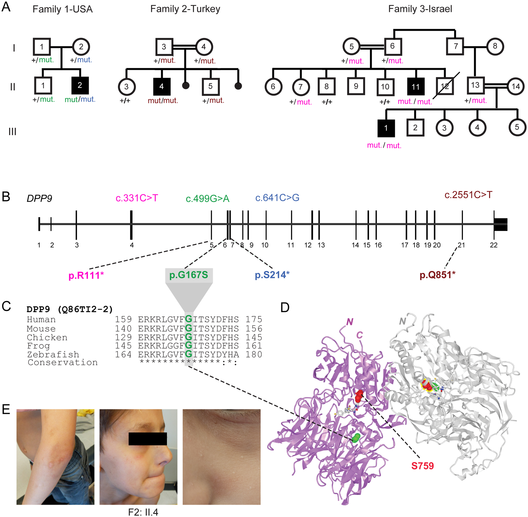
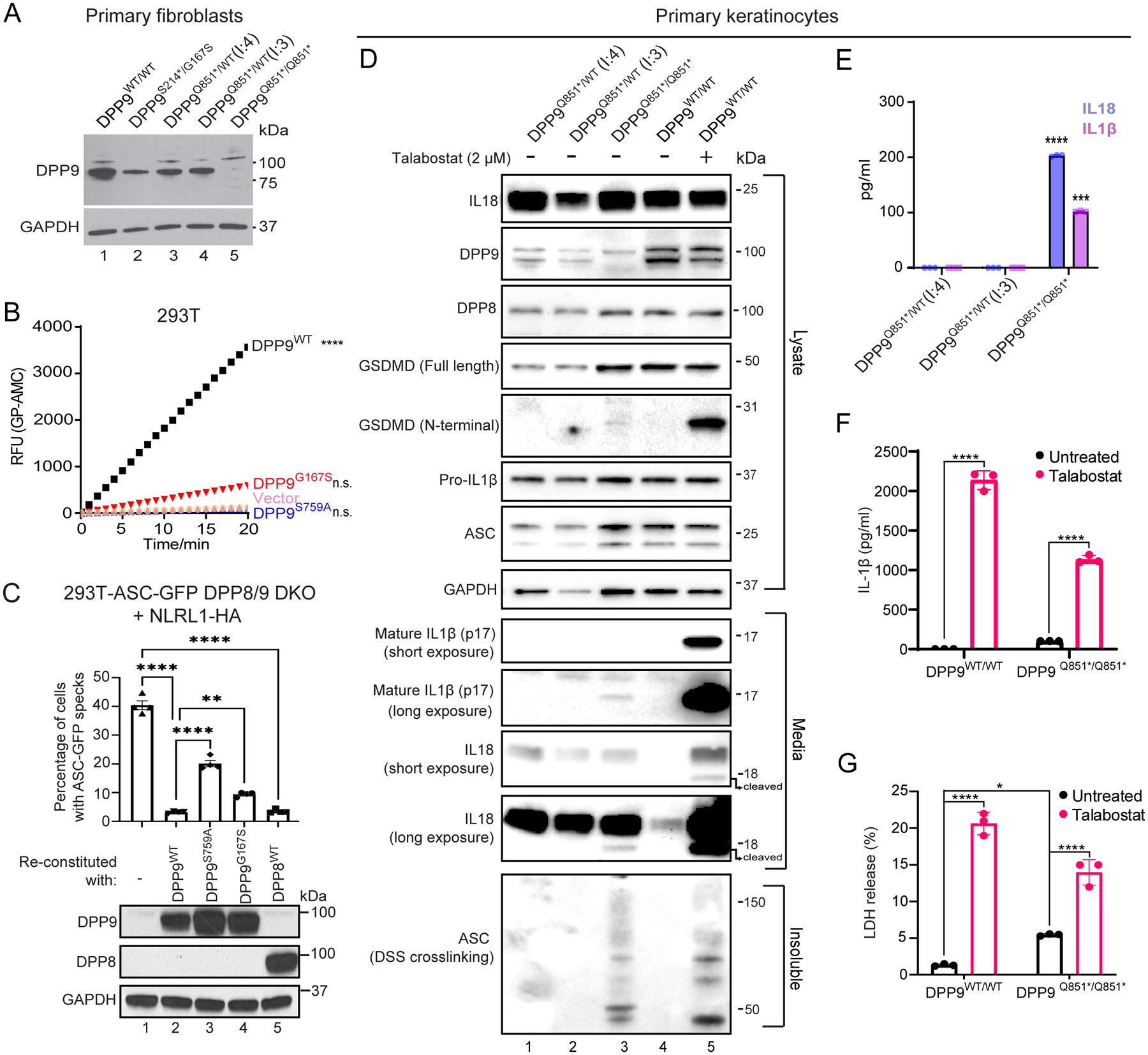
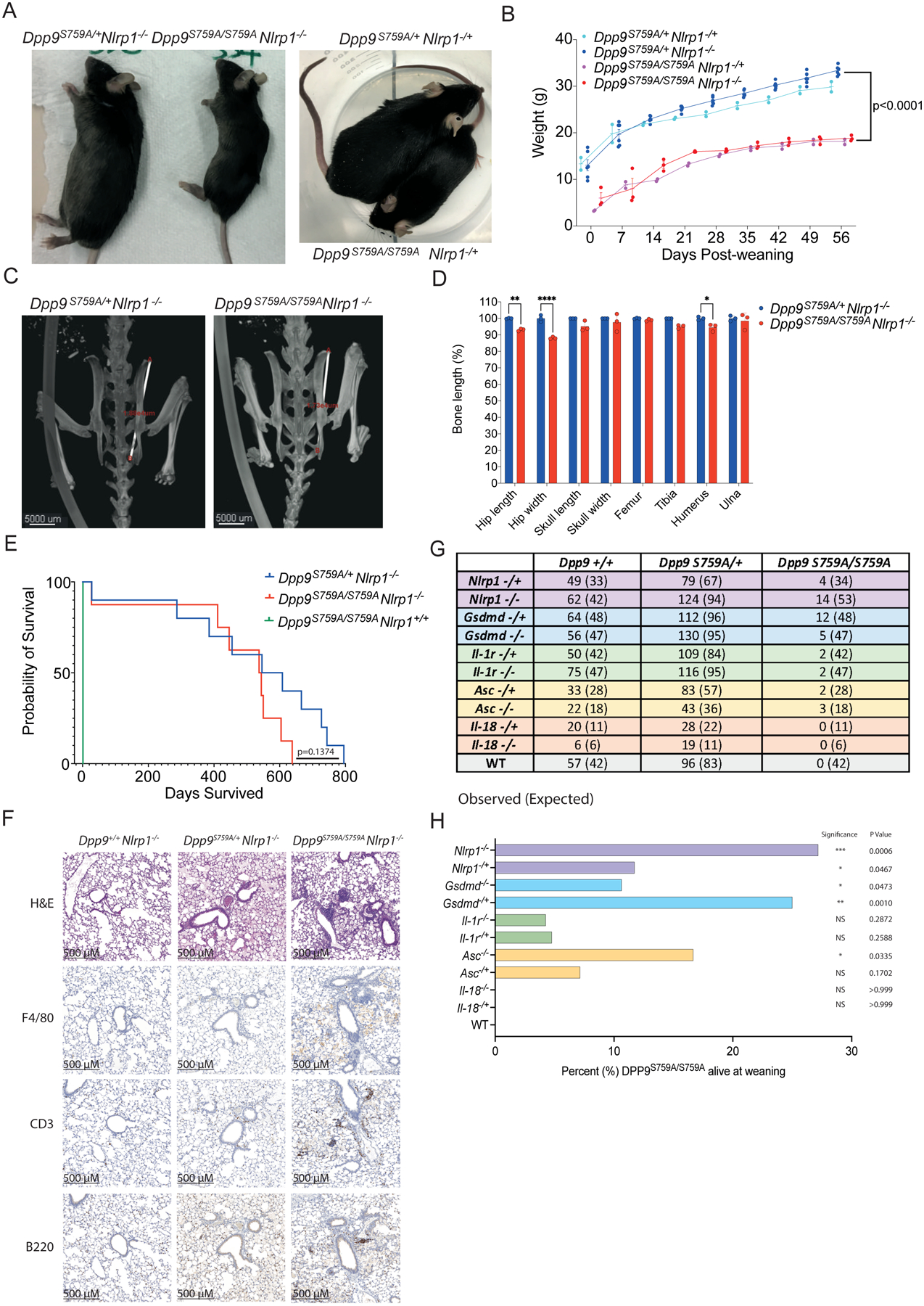
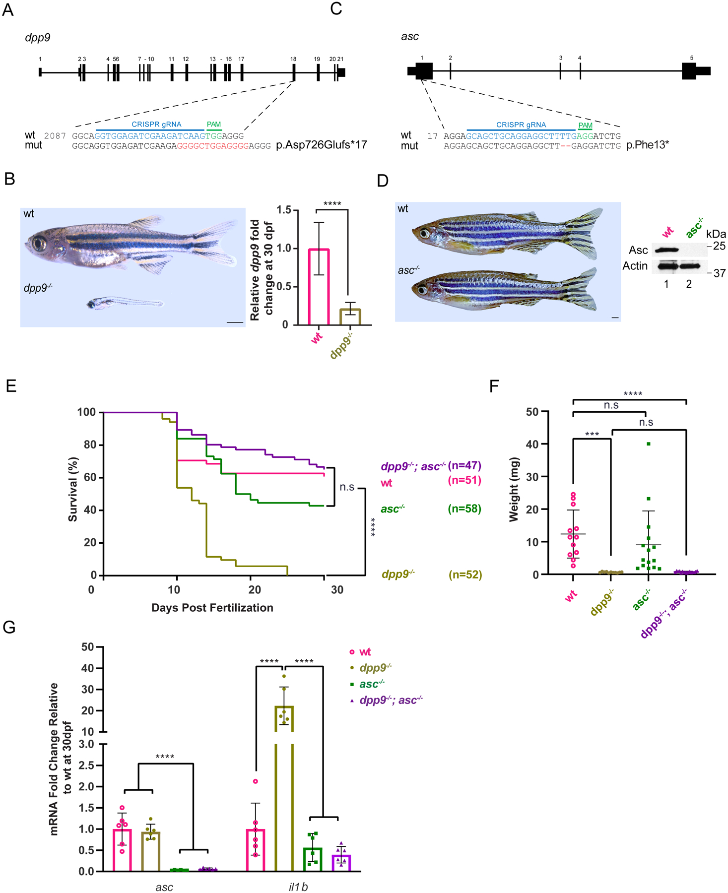
Dipeptidyl peptidase 9 (DPP9) is a direct inhibitor of NLRP1, but how it affects inflammasome regulation in vivo is not yet established. Here, we report three families with immune-associated defects, poor growth, pancytopenia, and skin pigmentation abnormalities that segregate with biallelic DPP9 rare variants. Using patient-derived primary cells and biochemical assays, these variants were shown to behave as hypomorphic or knockout alleles that failed to repress NLRP1. The removal of a single copy of Nlrp1a/b/c, Asc, Gsdmd, or Il-1r, but not Il-18, was sufficient to rescue the lethality of Dpp9 mutant neonates in mice. Similarly, dpp9 deficiency was partially rescued by the inactivation of asc, an obligate downstream adapter of the NLRP1 inflammasome, in zebrafish. These experiments suggest that the deleterious consequences of DPP9 deficiency were mostly driven by the aberrant activation of the canonical NLRP1 inflammasome and IL-1β signaling. Collectively, our results delineate a Mendelian disorder of DPP9 deficiency driven by increased NLRP1 activity as demonstrated in patient cells and in two animal models of the disease.


Conflict of interest statement

Competing interests:

S.L.M. is a Scientific Advisor for IFM Therapeutics and NRG Therapeutics.

Figures

Fig. 1:
Fig. 1:. Three families segregating recessive loss-of-function DPP9 germline variants
(A) Pedigrees of three families harboring germline DPP9 mutations. Proband from Family 1 (II.2, USA) is compound heterozygous, with paternal p.G167S and maternal p.S214* germline mutations. Proband from Family 2 (II.4, Turkey) carries a homozygous recessive p.Q851* mutation. Affected cousins from Family 3 (II.11 and III:1, Israel) carry a homozygous recessive p.R111* variant. (B) Exon-intron genomic organization of DPP9 with positions of identified germline mutations. (C) Phylogenetic conservation of the Gly167 residue, which is mutated in Family 1, across vertebrate species. (D) Protein structure of a DPP9 homodimer (PDB ID: 6EOR) depicting location of p.G167 on the periphery of the substrate binding site and p.S759 in the catalytic residue. (E) Photographs of proband from Family 2 (II.4) depicting recurrent HSV facial lesions, milia, and skin pigmentation anomalies on his arms.
Fig. 2:
Fig. 2:. Patients’ cells lack DPP9 or produce a strong enzymatic hypomorph
(A) Western blot analysis of DPP9 in primary dermal fibroblasts of an unrelated control (WT/WT) (lane 1), affected individual from family 1 (proband (II:2) S214*/G167S) (lane 2), two unaffected individuals from family 2 ((I:4) and (I:3) Q851*/WT) (lanes 3 and 4 respectively) and affected individual from family 2 (proband (II:4) Q851*/Q851*) (lane 5). (B) 293T cells were transfected with either vector, wild-type DPP9, DPP9 S759A or DPP9 G167S and lysed in PBS 1% Tween 20, 48 h after transfection. 0.3 μg of total lysate was then incubated with Gly-Pro-AMC fluorescence substrate. AMC fluorescence was measured for 20 mins at 25 °C in a 50 μl reaction every minute on a spectrometer. One-way ANOVA. ****P<0.0001 calculated by comparing the vector control to wild-type DPP9, DPP9S759A or DPP9G167S (n = 3 independent replicates). (C). Top, quantification of ASC-GFP speck formation in DPP8/9 DKO 293T-ASC-GFP cells reconstituted with WT DPP9, DPP9 S759A, DPP9 G167S or WT DPP8, and transfected with NLRP1. The percentage of cells with ASC-GFP specks were counted in three different fields after fixation. More than 100 cells were scored per condition. Data are mean ± SEM, n = 4 independent replicates. **p < 0.01; ****p < 0.0001. Bottom, western blot analysis validating DPP9 and DPP8 expression. (D) Western blot analysis of inflammasome components in primary keratinocytes of two unaffected individuals from family 2 ((I:4) and (I:3) Q851*/WT) and the affected individual from family 2 (proband (II:4) Q851*/Q851*). Additionally an unrelated control (+/+), treated with or without the potent DPP8/9 inhibitor Talabostat (2 μM) was included as a positive control for NLRP1 inflammasome activation. (E) IL1β and IL18 ELISA of the culture supernatant from primary keratinocytes from the two unaffected individuals from family 2 ((I:4) and (I:3) Q851*/WT) and affected individual from family 2 (proband (II:4) Q851*/Q851*). ****p < 0.001, ***p < 0.01 (one-way ANOVA). (n=3 replicates). (F), (G) Levels of IL-1β (F) and lactate dehydrogenase (LDH; G) released from DPP9WT/WT control and DPP9Q851*/Q851* patient-derived primary keratinocytes in absence or presence of Talabostat. Data are mean ± SEM, n = 3 independent replicates. *p < 0.05; ****p < 0.0001.
Fig. 3:
Fig. 3:. Deficiency of NLRP1 and downstream signaling molecules rescues neonatal lethality of Dpp9S759A/S759A mice
(A) Viable, 6-month-old male Dpp9S759A/S759A Nlrp1−/− and Dpp9S759A/S759A Nlrp1−/+ mice are runted compared to respective Dpp9S759A/+ Nlrp1−/− or Dpp9S759A/+ Nlrp1−/+ littermates. (B) Male Dpp9S759A/S759A Nlrp1−/− and Dpp9S759A/+ Nlrp1−/− mice were weighed weekly for 8 weeks post weaning. n=2–6 mice per genotype, error bars represent mean ±SD. Statistical significance was calculated based on the Dpp9S759A/S759A Nlrp1−/− and Dpp9S759A/+Nlrp1−/− mice using a paired t-test. (C) Example images of skeletal measurements and (D) quantification of 6-month-old Dpp9S759A/S759A Nlrp1−/− and Dpp9S759A/+ Nlrp1−/− mice imaged via MicroCT scanning. Measurements are normalised to the average Dpp9S759A/+ Nlrp1−/− values of mice of the same sex. n=3 mice per genotype, both male and female. Error bars represent mean ±SEM. *p<0.05, **p<0.005, ****p<0.0001 (2way ANOVA) (E) Kaplan Meier survival curve of Dpp9S759A/S759A Nlrp1−/− and Dpp9S759A/+Nlrp1−/− mice, starting at weaning age. n=8–13 per genotype, both male and female. Statistical significance was determined by a Mantel-Cox test. (F) Representative section of lung tissue from 6-month-old Dpp9S759A/S759A Nlrp1−/− and Dpp9S759A/+ Nlrp1−/− mice stained with Hematoxylin and eosin or immunohistochemistry of F4/80, B220 or CD3 antibodies. n=3 mice per genotype, both male and female. (G) The number of viable mice at weaning age (~3 weeks old) of various genotypes were genotyped and tallied, this is the ‘observed’ number depicted on the left of each cell. Mendelian ratios were used to calculate ‘Expected’ number of mice per genotype, written in brackets on the right of each cell. (H) Graphical representation of the percentage of Dpp9S759A/S759A mice surviving to weaning for each genetic cross. Statistical analysis was performed using Fisher’s exact test.
Fig. 4:
Fig. 4:. Deficiency of asc extends lifespan and abrogates pro-inflammatory il-1b upregulation in dpp9−/− zebrafish.
(A) Generation of dpp9 knockout zebrafish using CRISPR/Cas9-genome editing that resulted in an out-of-frame allele with premature stop codon. (B) Left, dpp9−/− zebrafish at 1 month of age exhibit a dramatic reduction in size (quantification shown in F). Right, validation of dpp9-KO by RT-qPCR. Data are mean ± SEM (n = 6). ****p<0.001 (one-way ANOVA). (C) Generation of asc knockout zebrafish using CRISPR/Cas9 genome editing that resulted in a 2-bp deletion in exon 1 followed by a premature stop codon. (D) Left, asc−/− zebrafish appeared indistinguishable from wt. Right, validation of complete asc-KO by western blotting. (E) Kaplan-Meier survival curves of wt, dpp9−/−, asc−/− and double dpp9−/−; asc−/− zebrafish. Dpp9−/− zebrafish demonstrated a significant survival disadvantage that is rescued by inactivating asc. ****p<0.0001 (Logrank Mantel-Cox test). (F) The weight of double dpp9−/−; asc−/− zebrafish is not rescued compared to that of dpp9−/− zebrafish. Data are mean ± SEM. ****p<0.001; ***p<0.01; n.s., non-significant (one-way ANOVA). (G) In double dpp9−/−; asc−/− zebrafish, il1b expression is rescued to that of wt levels as demonstrated by RT-qPCR analysis. Data are mean ± SEM (n = 6). ****p<0.001 (one-way ANOVA).

References

    1. Robinson KS, Teo DET, Tan KS, Toh GA, Ong HH, Lim CK, Lay K, Au BV, Lew TS, Chu JJH, Chow VTK, Wang DY, Zhong FL, Reversade B, Enteroviral 3C protease activates the human NLRP1 inflammasome in airway epithelia. Science. 370 (2020), doi: 10.1126/science.aay2002. - DOI - PubMed
    1. Bauernfried S, Scherr MJ, Pichlmair A, Duderstadt KE, Hornung V, Human NLRP1 is a sensor for double-stranded RNA. Science (2020), doi: 10.1126/science.abd0811. - DOI - PubMed
    1. Fenini G, Grossi S, Contassot E, Biedermann T, Reichmann E, French LE, Beer H-D, Genome Editing of Human Primary Keratinocytes by CRISPR/Cas9 Reveals an Essential Role of the NLRP1 Inflammasome in UVB Sensing. J. Invest. Dermatol 138, 2644–2652 (2018). - PubMed
    1. Broz P, Dixit VM, Inflammasomes: mechanism of assembly, regulation and signalling. Nat. Rev. Immunol 16, 407–420 (2016). - PubMed
    1. Zhong FL, Mamaï O, Sborgi L, Boussofara L, Hopkins R, Robinson K, Szeverényi I, Takeichi T, Balaji R, Lau A, Tye H, Roy K, Bonnard C, Ahl PJ, Jones LA, Baker PJ, Lacina L, Otsuka A, Fournie PR, Malecaze F, Lane EB, Akiyama M, Kabashima K, Connolly JE, Masters SL, Soler VJ, Omar SS, McGrath JA, Nedelcu R, Gribaa M, Denguezli M, Saad A, Hiller S, Reversade B, Germline NLRP1 Mutations Cause Skin Inflammatory and Cancer Susceptibility Syndromes via Inflammasome Activation. Cell. 167, 187–202.e17 (2016). - PubMed

MeSH terms