. 2022 Nov 15;82(22):4288-4298.
doi: 10.1158/0008-5472.CAN-21-2483.

A Probody T Cell-Engaging Bispecific Antibody Targeting EGFR and CD3 Inhibits Colon Cancer Growth with Limited Toxicity

Affiliations

A Probody T Cell-Engaging Bispecific Antibody Targeting EGFR and CD3 Inhibits Colon Cancer Growth with Limited Toxicity

Leila M Boustany et al. Cancer Res. .

Abstract

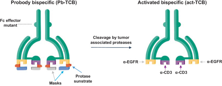
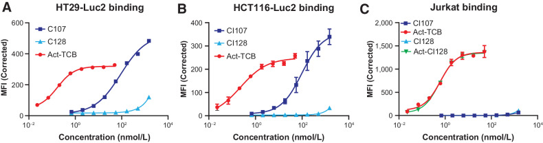
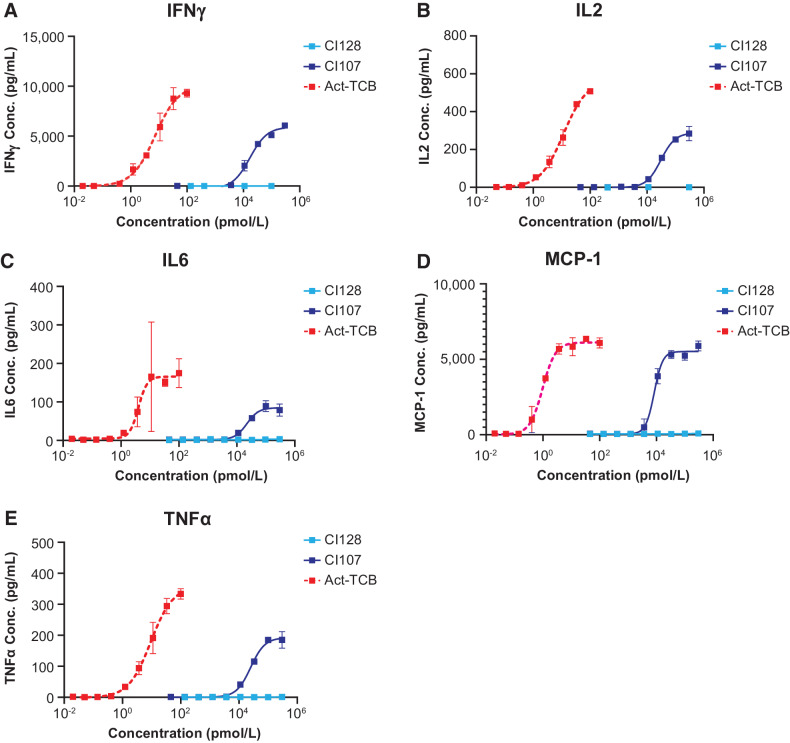
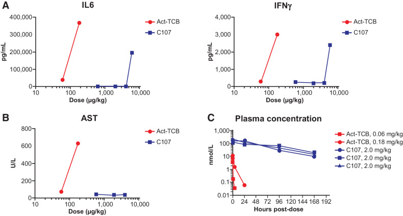
T cell-engaging bispecific antibodies (TCB) are highly potent therapeutics that can recruit and activate cytotoxic T cells to stimulate an antitumor immune response. However, the development of TCBs against solid tumors has been limited by significant on-target toxicity to normal tissues. Probody therapeutics have been developed as a novel class of recombinant, protease-activated antibody prodrugs that are "masked" to reduce antigen binding in healthy tissues but can become conditionally unmasked by proteases that are preferentially active in the tumor microenvironment (TME). Here, we describe the preclinical efficacy and safety of CI107, a Probody TCB targeting EGFR and CD3. In vitro, the protease-activated, unmasked CI107 effectively bound EGFR and CD3 expressed on the surface of cells and induced T-cell activation, cytokine release, and cytotoxicity toward tumor cells. In contrast, dually masked CI107 displayed a >500-fold reduction in antigen binding and >15,000-fold reduction in cytotoxic activity. In vivo, CI107 potently induced dose-dependent tumor regression of established colon cancer xenografts in mice engrafted with human peripheral blood mononuclear cells. Furthermore, the MTD of CI107 in cynomolgus monkeys was more than 60-fold higher than that of the unmasked TCB, and much lower levels of toxicity were observed in animals receiving CI107. Therefore, by localizing activity to the TME and thus limiting toxicity to normal tissues, this Probody TCB demonstrates the potential to expand clinical opportunities for TCBs as effective anticancer therapies for solid tumor indications.

Significance: A conditionally active EGFR-CD3 T cell-engaging Probody therapeutic expands the safety window of bispecific antibodies while maintaining efficacy in preclinical solid tumor settings.


Figures

Figure 1. Design of Probody T cell–engaging bispecific therapeutic (Probody TCB). CI107 comprises a bispecific IgG antibody with EGFR and CD3 binding domains. Peptide masks of both binding domains are attached via a protease-cleavable linker. In normal tissues, these masks are designed to prevent binding to the target protein and to CD3+ lymphocytes. In contrast, cleavage of the linker by proteases present in the solid tumor microenvironment expose the EGFR and CD3 binding domains, enabling specific binding of the Probody TCB to the target antigen and CD3+ T cells.
Figure 1.
Design of Probody T cell–engaging bispecific therapeutic (Probody TCB). CI107 comprises a bispecific IgG antibody with EGFR and CD3 binding domains. Peptide masks of both binding domains are attached via a protease-cleavable linker. In normal tissues, these masks are designed to prevent binding to the target protein and to CD3+ lymphocytes. In contrast, cleavage of the linker by proteases present in the solid tumor microenvironment expose the EGFR and CD3 binding domains, enabling specific binding of the Probody TCB to the target antigen and CD3+ T cells.
Figure 2. Dual masking of the anti-EGFR and anti-CD3 binding domains attenuates binding of CI107 to EGFR and CD3 expressed on the surface of cells. HT29 (A) HCT116 (B), or Jurkat (C) cells were incubated with increasing concentrations of CI107 or protease-activated TCB (Act-TCB), and binding was assessed by flow cytometry.
Figure 2.
Dual masking of the anti-EGFR and anti-CD3 binding domains attenuates binding of CI107 to EGFR and CD3 expressed on the surface of cells. HT29 (A), HCT116 (B), or Jurkat (C) cells were incubated with increasing concentrations of CI107 or protease-activated TCB (Act-TCB), and binding was assessed by flow cytometry.
Figure 3. Masking of EGFR and CD3 binding domains attenuates cytotoxicity and T-cell activation mediated by CI107 in PBMCs cocultured with human colorectal cancer cells. HCT116-Luc2 (A, C) and HT29-Luc2 (B, D) cells were cocultured with human PBMCs (Donor 4) and treated with increasing concentrations of CI107, activated TCB (Act-TCB), or non-EGFR-targeted control CI128. (A, B) After 48 hours of culture, HCT116-Luc2 or HT29-Luc2 cell viability was measured by the ONE-Glo Luciferase Assay, and cytotoxicity was measured relative to untreated controls. C and D, After 16 hours of culture, CD69 expression in PBMCs was measured by flow cytometry. MFI, mean fluorescence intensity.
Figure 3.
Masking of EGFR and CD3 binding domains attenuates cytotoxicity and T-cell activation mediated by CI107 in PBMCs cocultured with human colorectal cancer cells. HCT116-Luc2 (A and C) and HT29-Luc2 (B and D) cells were cocultured with human PBMCs (Donor 4) and treated with increasing concentrations of CI107, activated TCB (Act-TCB), or non-EGFR-targeted control CI128. A and B, After 48 hours of culture, HCT116-Luc2 or HT29-Luc2 cell viability was measured by the ONE-Glo Luciferase Assay, and cytotoxicity was measured relative to untreated controls. C and D, After 16 hours of culture, CD69 expression in PBMCs was measured by flow cytometry. MFI, mean fluorescence intensity.
Figure 4. Masking of EGFR and CD3 binding domains in CI107 attenuates cytokine release in PBMCs cocultured with human colorectal cancer cells. HCT116-Luc2 cells were cocultured with human PBMCs (Donor 4) and treated with increasing concentrations of CI107, activated TCB (Act-TCB), or non–EGFR-targeted control CI128. After 16 hours of culture, cytokine release was measured for IFNγ (A), IL2 (B), IL6 (C), MCP-1 (D), and TNFα (E) using a U-PLEX plate.
Figure 4.
Masking of EGFR and CD3 binding domains in CI107 attenuates cytokine release in PBMCs cocultured with human colorectal cancer cells. HCT116-Luc2 cells were cocultured with human PBMCs (Donor 4) and treated with increasing concentrations of CI107, activated TCB (Act-TCB), or non–EGFR-targeted control CI128. After 16 hours of culture, cytokine release was measured for IFNγ (A), IL2 (B), IL6 (C), MCP-1 (D), and TNFα (E) using a U-PLEX plate.
Figure 5. Sensitivity of Probody TCB to protease cleavage correlates with efficacy and intratumoral T-cell presence in PBMC-engrafted NSG mice. A, NSG mice harboring HT29-Luc2 tumors and engrafted with human PBMCs were treated on days 1, 8, and 15 with vehicle (PBS) or 0.3 mg/kg CI020, CI011, CI040, or CI048 (n = 8 per group). Tumor volume was measured twice weekly. B, NSG mice harboring HT29-Luc2 tumors and engrafted with human PBMCs were treated with vehicle or 1 mg/kg of CI020, CI011, CI040, or CI048. Tumors were harvested 7 days after dosing, and immunohistochemistry for CD3 was performed. Brown stain indicates CD3+ cells.
Figure 5.
Sensitivity of Probody TCB to protease cleavage correlates with efficacy and intratumoral T-cell presence in PBMC-engrafted NSG mice. A, NSG mice harboring HT29-Luc2 tumors and engrafted with human PBMCs were treated on days 1, 8, and 15 with vehicle (PBS) or 0.3 mg/kg CI020, CI011, CI040, or CI048 (n = 8 per group). Tumor volume was measured twice weekly. B, NSG mice harboring HT29-Luc2 tumors and engrafted with human PBMCs were treated with vehicle or 1 mg/kg of CI020, CI011, CI040, or CI048. Tumors were harvested 7 days after dosing, and immunohistochemistry for CD3 was performed. Brown stain indicates CD3+ cells.
Figure 6. CI107 induces dose-dependent regression of xenograft tumors in PBMC-engrafted NSG mice. NSG mice harboring HT29 (A) or HCT116 (B) xenograft tumors and engrafted with human PBMCs were treated with CI107 once weekly for 3 weeks. Tumor volume was measured twice weekly. *, P < 0.5; **, P < 0.01; ****, P < 0.0001.
Figure 6.
CI107 induces dose-dependent regression of xenograft tumors in PBMC-engrafted NSG mice. NSG mice harboring HT29 (A) or HCT116 (B) xenograft tumors and engrafted with human PBMCs were treated with CI107 once weekly for 3 weeks. Tumor volume was measured twice weekly. *, P < 0.5; **, P < 0.01; ****, P < 0.0001.
Figure 7. CI107 provides improved tolerability and exposure relative to Act-TCB in cynomolgus monkeys. Cynomolgus monkeys were administered a single dose of 0.06 or 0.18 mg/kg of activated TCB (Act-TCB) on day 1 or 0.6, 2.0, 4.0, or 6.0 mg/kg CI107. A, Levels of IL6 and IFNγ were measured 8 hours after dosing. Cytokine analysis was performed with a Luminex suspension array system on serum samples. B, Levels of AST were measured by serum chemistry analysis 48 hours after dosing. C, Plasma concentrations of Act-TCB and CI107 were measured by ELISA using anti-idiotype capture and anti-human Fc detection. Blue lines in C represent data from 3 individual animals dosed with 2.0 mg/kg CI107; red lines represent single animals dosed with 0.06 or 0.18 mg/kg Act-TCB.
Figure 7.
CI107 provides improved tolerability and exposure relative to Act-TCB in cynomolgus monkeys. Cynomolgus monkeys were administered a single dose of 0.06 or 0.18 mg/kg of activated TCB (Act-TCB) on day 1 or 0.6, 2.0, 4.0, or 6.0 mg/kg CI107. A, Levels of IL6 and IFNγ were measured 8 hours after dosing. Cytokine analysis was performed with a Luminex suspension array system on serum samples. B, Levels of AST were measured by serum chemistry analysis 48 hours after dosing. C, Plasma concentrations of Act-TCB and CI107 were measured by ELISA using anti-idiotype capture and anti-human Fc detection. Blue lines in C represent data from three individual animals dosed with 2.0 mg/kg CI107; red lines represent single animals dosed with 0.06 or 0.18 mg/kg Act-TCB.

References

    1. Kantarjian H, Stein A, Gokbuget N, Fielding AK, Schuh AC, et al. . Blinatumomab versus chemotherapy for advanced acute lymphoblastic leukemia. N Engl J Med 2017;376:836–47. - PMC - PubMed
    1. Gökbuget N, Dombret H, Bonifacio M, Reichle A, Graux C, Faul C, et al. . Blinatumomab for minimal residual disease in adults with B-cell precursor acute lymphoblastic leukemia. Blood 2018;131:1522–31. - PMC - PubMed
    1. Guy DG, Uy GL. Bispecific antibodies for the treatment of acute myeloid leukemia. Curr Hematol Malig Rep 2018;13:417–25. - PMC - PubMed
    1. Caraccio C, Krishna S, Phillips DJ, Schürch CM. Bispecific antibodies for multiple myeloma: a review of targets, drugs, clinical trials, and future directions. Front Immunol 2020;11:501. - PMC - PubMed
    1. Trabolsi A, Arumov A, Schatz JH. T cell-activating bispecific antibodies in cancer therapy. J Immunol 2019;203:585–92. - PubMed

Publication types

MeSH terms