

Wide Arterial Sparing Encephalo-Duro-Synangiosis for Moyamoya: Surgical Technique and Outcomes
- PMID: 36113163
- PMCID: PMC10593263
- DOI: 10.1227/ons.0000000000000376
Wide Arterial Sparing Encephalo-Duro-Synangiosis for Moyamoya: Surgical Technique and Outcomes
Erratum in
-
Wide Arterial Sparing Encephalo-Duro-Synangiosis for Moyamoya: Surgical Technique and Outcomes: Corrigendum.Oper Neurosurg. 2023 Apr 1;24(4):e314-314. doi: 10.1227/ons.0000000000000646. Oper Neurosurg. 2023. PMID: 36920059 Free PMC article. No abstract available.
Abstract
Background: Moyamoya is managed by surgical revascularization, but no standardized method has yet been universally adopted.
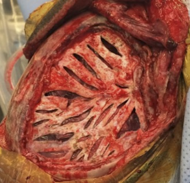
Objective: To describe a new indirect bypass technique for pediatric moyamoya, wide arterial sparing encephalo-duro-synangiosis (WASEDS), which provides a much wider area of revascularization with minimal compromise to the middle meningeal arterial tree compared with traditional procedures. Initially used as a salvage technique after failed encephalo-duro-arterio-synangiosis, its success later motivated its use as a first-line procedure.
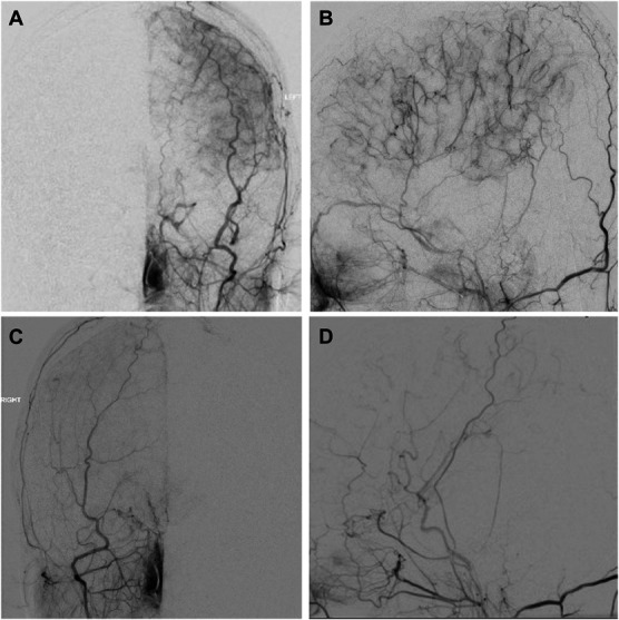
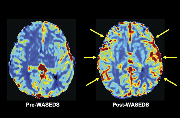
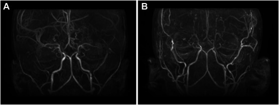
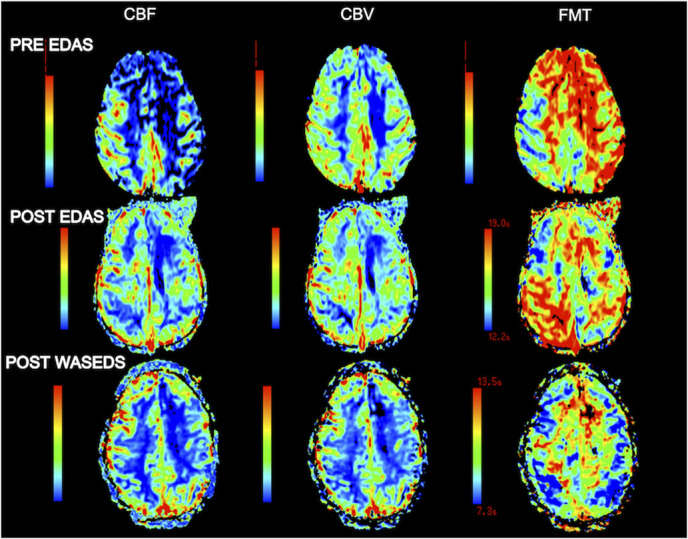
Methods: Clinical and radiographic records of patients who underwent WASEDS for moyamoya from 2009 to 2020 were reviewed. Brain perfusion relative cerebral blood volume on the side of the WASEDS procedure was calculated. Two-tailed paired t tests were performed to identify the statistically significant differences ( P ≤ .05).
Results: WASEDS was successfully performed on 8 patients for a total of 14 cerebral hemispheres. Age ranged from 2 to 25 years. There were no mortalities. The average clinical and radiographic follow-up was 49.79 months (range 2-126 months), demonstrating improvement in neurological condition and no postoperative stroke and significant diminution or cessation of transient ischemic attacks in all patients. Relative cerebral blood volume increased 9.24% after the WASEDS procedure ( P = .012). There were no neurological complications. There were 2 pseudomeningoceles related to the extensive dural openings.
Conclusion: WASEDS is a safe and effective indirect revascularization technique for both primary and salvage techniques. It provides an extensive area of cortical revascularization with no compromise of the middle meningeal vasculature and subjective reports of early improvement in cognition and behavior. The main disadvantage is elevated risk of pseudomeningocele secondary to the large craniotomy.
Copyright © Congress of Neurological Surgeons 2022. All rights reserved.
Figures
Comment in
-
Letter: Wide Arterial Sparing Encephalo-Duro-Synangiosis for Moyamoya: Surgical Technique and Outcomes.Oper Neurosurg. 2023 May 1;24(5):e391. doi: 10.1227/ons.0000000000000681. Epub 2023 Mar 15. Oper Neurosurg. 2023. PMID: 36921246 No abstract available.
References
-
- Jodi LS. Understanding and treating moyamoya disease in children. Neurosurg Focus. 2009;26(4):1-11. - PubMed
-
- Fung LWE, Thompson D, Ganesan V. Revascularisation surgery for paediatric moyamoya: a review of the literature. Childs Nerv Syst. 2005;21(5):358-364. - PubMed
-
- Scott RM, Smith JL, Robertson RL, Madsen JR, Soriano SG, Rockoff MA. Long-term outcome in children with moyamoya syndrome after cranial revascularization by pial synangiosis. J Neurosurg. 2004;100(2 suppl):142-149. - PubMed
-
- Matsushima Y, Inaba Y. The specificity of the collaterals to the brain through the study and surgical treatment of moyamoya disease. Stroke. 1986;17(1):117-122. - PubMed
Publication types
MeSH terms
Grants and funding
LinkOut - more resources
Full Text Sources
