. 2023 Feb;17(1):6-16.
doi: 10.14444/8357. Epub 2022 Sep 16.

Anterior Vertebral Body Tethering for Scoliosis Patients With and Without Skeletal Growth Remaining: A Retrospective Review With Minimum 2-Year Follow-Up

Affiliations

Anterior Vertebral Body Tethering for Scoliosis Patients With and Without Skeletal Growth Remaining: A Retrospective Review With Minimum 2-Year Follow-Up

Theodor Di Pauli von Treuheim et al. Int J Spine Surg. 2023 Feb.

Abstract

Background: Anterior vertebral body tethering (AVBT) has been approved for skeletally immature (IM) adolescent idiopathic scoliosis patients, but the role of AVBT in patients with minimal remaining skeletal growth is controversial. The purpose of this study was to compare minimum 2-year (YR2) outcomes in skeletally IM patients vs those with minimal remaining skeletal growth.

Methods: Patients with single thoracic AVBT were grouped by their preoperative (PR) skeletal maturity: IM (n = 16, Risser 0-2) vs mature (M, n = 19, Risser 3-5). Outcomes were assessed at PR, first erect (FE), and YR2. Median (range) was compared with nonparametric tests (P < 0.05).

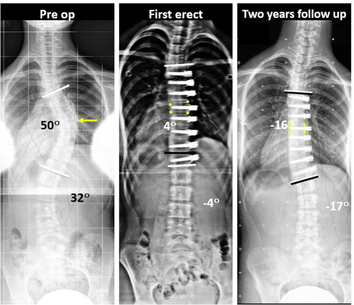
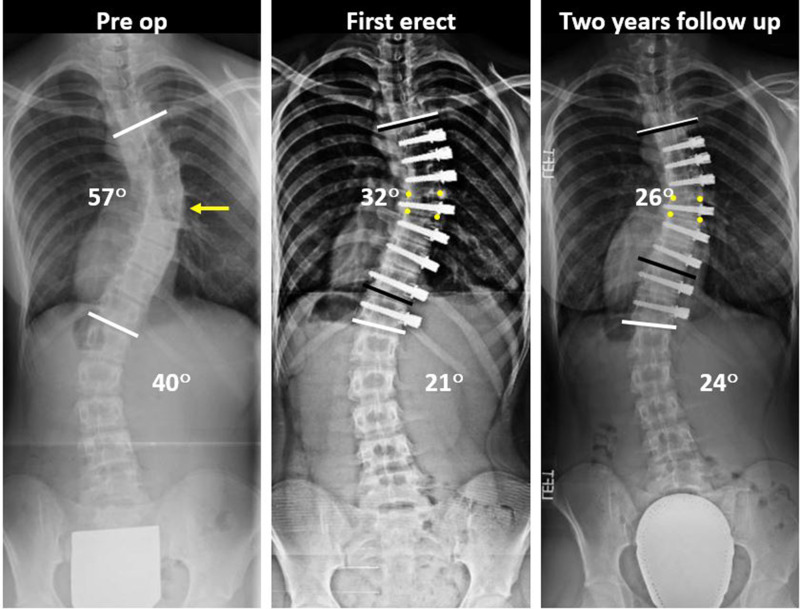
Results: The PR age was 12.5 (9-16) vs 15 (12-18) years with major Cobb 51° (36°-69°) and 49° (40°-69°) for IM and M, respectively. At FE, there was no difference in correction; however, at YR2, the IM group yielded a lower residual curve (15° [-16° to 38°] vs 29° [12°-42°], P = 0.008). Thoracolumbar/lumbar curves were corrected without group differences. Clinically successful correction (<35°) (15 [94%] vs 15 [79%]) and suspected cord breakages (2 [13%] vs 2 [12%]) were similar at YR2. Two overcorrections occurred, both in IM patients. Scoliosis Research Society-22 outcomes at final follow-up were similar between groups. No revision reoperations or conversions to spinal fusion were needed.

Conclusions: Skeletally IM patients benefit from greater growth-modulated curve correction than M patients, however, at the increased risk of overcorrection. M patients maintained clinically significant correction at latest follow-up. Longer-term follow-up is required to determine durability of outcomes for patients undergoing AVBT who have minimal remaining growth at the time of index surgery.

Clinical relevance: This study is relevant to spine surgeons, spine physiotherapists, and patients with idiopathic scoliosis. It offers evidence of clinical correction of scoliosis in mature patients.

Keywords: adolescent idiopathic scoliosis; non-fusion surgery; scoliosis; vertebral body tethering.


Conflict of interest statement

Declaration of Conflicting Interests: Dr Lonner reports personal fees, royalty fees, and research grant support from Zimmer Biomet for The Tether implant. Dr Lonner also reports personal fees, nonfinancial support and other from Depuy Synthes, personal fees and non-financial support from OrthoPediatrics, other from Paradigm Spine, non-financial support and other from Spine Search, and other from Setting Scoliosis Straight Foundation outside the submitted work.

Figures

Figure 1
Figure 1
Sagittal curve behavior, characterized as T5-T12 kyphosis, shows an inverse relationship between preoperative kyphosis and the change in curvature over 2 y. This relationship was statistically significant for immature patients whose spines are more amenable to growth modulation from anterolateral compression.
Figure 2
Figure 2
Coronal and sagittal radiographic measurements at 3 timepoints show differences due to skeletal maturity only at 2-y follow-up in the instrumented thoracic curve. Differences between groups are designated by black bar. Differences within a group with reference to preoperative and first erect values are denoted by asterisk (*) and cross (‡), respectively. Boxplot interquartile range shows 25% to 75% with black bar indicating median. Outliers are designated by red plus sign (1.5 times interquartile range). Between- and within-group comparisons were done using a Mann-Whitney U test with Bonferroni correction (P < 0.05). PR, preoperative; FE, first erect; YR2, 2 y; T, thoracic; TL/L, thoracolumbar/lumbar.
Figure 3
Figure 3
Major and compensatory curve first erect and total percent correction (FE% and YR2%) are referenced to preoperative (PR) curve, whereas follow-up percent correction (FU%) compares FE with YR2. Percent correction beyond 100% defines overcorrection, and negative values define loss of correction. Large variations seen in FU% occurs with near-zero FE curves. Boxplots follow formatting described in Figure 2. Between-group comparisons were done using a Mann-Whitney U test with Bonferroni correction (P < 0.05).
Figure 4
Figure 4
(A) Vertical height changes of the instrumented segment show significant differences between groups at preoperative (PR) and first erect (FE) that disappear at 2 y (YR2). (B) The immature group had greater height gain at YR2, but this difference disappeared when normalizing by total body height gain. Between- and within-group comparisons were done using a Mann-Whitney U test with Bonferroni correction (P < 0.05). UIV-LIV, upper and lower instrumented levels.
Figure 5
Figure 5
(A) The degree of apical vertebral body wedging was assessed at first erect (FE) and compared with the 2-y (YR2) wedge. (B) Vertebral body squaring (VBSQ) quantifies the difference in wedging from FE to YR2, where more positive values indicate greater squaring. Immature patients showed significantly more squaring. (c) Representative immature and mature patients highlight changes in wedging. Between-group comparisons were done using a Mann-Whitney U test with Bonferroni correction (P < 0.05).
Figure 6
Figure 6
Correction of major thoracic (instrumented) and compensatory thoracolumnbar/lumbar (noninstrumented) curves toward target region (light blue, <35°) is captured from preoperative (PR) (unfilled) and first erect (FE) (unfilled) to 2-y (YR2) follow-up (filled). Overcorrection of major (Q1), compensatory (Q3), or both curves (Q2) is highlighted in orange. Suspected breakage is indicated by star.
Figure 7
Figure 7
Preoperative posteroanterior scoliosis radiograph of an immature group patient (Risser 2, proximal humerus ossification system 1 [PHOS 1]) with a 50° thoracic curve and 32° thoracolumbar curve. First erect (FE) radiograph shows correction of major and compensatory curves. FE% correction was 92%. The patient was Risser 4 (PHOS 5) at 24 mo, and posteroanterior radiograph shows major curve overcorrection (2-y percent correction [YR2%] = 132%), corresponding to a 500% follow-up percent change (from FE to YR2). Apical wedge angle was 8° at FE and 0° at YR2, generating a vertebral body squaring of 8°. Follow-up angles were measured between vertebra contained by the black bars, where white bars indicate preoperative angles. Yellow arrow indicates the apical vertebra, and yellow dots mark the vertebral body margins.
Figure 8
Figure 8
Preoperative posteroanterior scoliosis radiograph of a mature group patient (Risser 4, proximal humerus ossification system 4 [PHOS 4]) with a 57° thoracic curve and 40° lumbar curve. First erect radiograph shows correction of major and compensatory curves. At 24 mo, the patient was Risser 5 (PHOS 5), and posteroanterior radiograph shows maintenance of clinically successful correction. Apical wedge angle was 12° at first erect and 8° at 2-y follow-up, calculating a VBSQ of 4°. Follow-up Cobb angles were measured between vertebra contained by the black bars, where white bars indicate preoperative angles. The yellow arrow indicates the apical vertebra, and yellow dots mark the vertebral body margins.

References

    1. Mente PL, Stokes IA, Spence H, Aronsson DD. Progression of vertebral wedging in an asymmetrically loaded rat tail model. Spine (Phila Pa 1976). 1997;22(12):1292–1296. 10.1097/00007632-199706150-00003 - DOI - PubMed
    1. Stokes IA, Spence H, Aronsson DD, Kilmer N. Mechanical modulation of vertebral body growth. Implications for scoliosis progression. Spine (Phila Pa 1976). 1996;21(10):1162–1167. 10.1097/00007632-199605150-00007 - DOI - PubMed
    1. Weinstein SL, Dolan LA, Cheng JCY, Danielsson A, Morcuende JA. Adolescent idiopathic scoliosis. Lancet. 2008;371(9623):1527–1537. 10.1016/S0140-6736(08)60658-3 - DOI - PubMed
    1. Danielsson AJ, Romberg K, Nachemson AL. Spinal range of motion, muscle endurance, and back pain and function at least 20 years after fusion or brace treatment for adolescent idiopathic scoliosis: a case-control study. Spine (Phila Pa 1976). 2006;31(3):275–283. 10.1097/01.brs.0000197652.52890.71 - DOI - PubMed
    1. Hoernschemeyer DG, Boeyer ME, Robertson ME, et al. . Anterior vertebral body tethering for adolescent scoliosis with growth remaining: a retrospective review of 2 to 5-year postoperative results. J Bone Joint Surg Am. 2020;102(13):1169–1176. 10.2106/JBJS.19.00980 - DOI - PubMed