. 2022 Sep 29;9(5):ENEURO.0269-22.2022.
doi: 10.1523/ENEURO.0269-22.2022. Print 2022 Sep-Oct.

Ventrolateral Prefrontal Cortex Contributes to Human Motor Learning

Affiliations

Ventrolateral Prefrontal Cortex Contributes to Human Motor Learning

Neeraj Kumar et al. eNeuro. .

Abstract

This study assesses the involvement in human motor learning, of the ventrolateral prefrontal cortex (BA 9/46v), a somatic region in the middle frontal gyrus. The potential involvement of this cortical area in motor learning is suggested by studies in nonhuman primates which have found anatomic connections between this area and sensorimotor regions in frontal and parietal cortex, and also with basal ganglia output zones. It is likewise suggested by electrophysiological studies which have shown that activity in this region is implicated in somatic sensory memory and is also influenced by reward. We directly tested the hypothesis that area 9/46v is involved in reinforcement-based motor learning in humans. Participants performed reaching movements to a hidden target and received positive feedback when successful. Before the learning task, we applied continuous theta burst stimulation (cTBS) to disrupt activity in 9/46v in the left or right hemisphere. A control group received sham cTBS. The data showed that cTBS to left 9/46v almost entirely eliminated motor learning, whereas learning was not different from sham stimulation when cTBS was applied to the same zone in the right hemisphere. Additional analyses showed that the basic reward-history-dependent pattern of movements was preserved but more variable following left hemisphere stimulation, which suggests an overall deficit in somatic memory for target location or target directed movement rather than reward processing per se. The results indicate that area 9/46v is part of the human motor learning circuit.

Keywords: TMS; motor learning; reinforcement.


Figures

Figure 1.
Figure 1.
Participants learned to make movements to a hidden target, and positive feedback was provided for successful movements. A, Participants made movements holding a robotic manipulandum. B, Schematic of the task. Participants made outward movements. If the movement direction fell within the hidden target zone, positive feedback was provided to indicate success. No feedback was given in the case of an unsuccessful movement. C, Experimental sequence. MEPs were elicited from the motor hot-spot in the left or right hemisphere before stimulation (cTBS to right or left 9/46v or sham stimulation). MEPs were again recorded 10 min after stimulation followed by the motor learning trials. In the no-feedback session at the end, participants were not provided with feedback on the success of the movement. D, Location of the stimulation site in representative participants from the left 9/46v and right 9/46v condition, shown in the sagittal (right panel) and coronal (middle panel) planes. The average location of the stimulation site (red circle) across participants in the MNI brain.
Figure 2.
Figure 2.
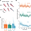
Suppression of left 9/46v using cTBS disrupts motor learning. A, Hand paths of a representative participant from each group at the start (block 1) and end of training (block 4). Hand paths shown in red are for unsuccessful movements, and those in blue are for successful movements. B, Mean absolute deviation from the center of the target zone over the course of training. The linear fit is shown across learning trials and no-feedback trials separately. The shaded region represents ±SEM. The rate of learning was less in participants who received stimulation over left 9/46v than those who received sham stimulation. C, Mean absolute deviation in the first and last block of the training. Participants in the sham stimulation condition showed a greater reduction in |AD| than participants in the left 9/46v condition.
Figure 3.
Figure 3.
Suppression of left 9/46v using cTBS leaves reinforcement learning intact. A, Mean percentage of rewarded trials over the course of training. A linear fit is shown across learning trials. The shaded region represents ±SEM. B, Mean percent of rewarded movements in the first and last block of the training. Participants in the sham stimulation condition received more rewards as learning progressed, whereas participants who received stimulation to left 9/46v showed no improvement at all. C, Mean absolute change in movement direction between the current trial (nth trial) and the subsequent trial (n + 1th trial) as a function of the history of rewarded movements. Reward history included three most recent movements (n, n–1, and n–2 trial), where at least one of these movements was rewarded. The left 9/46v group showed the same basic reward-history-dependent pattern as the other conditions but with greater change in direction overall. This suggests that the learning deficit after left 9/46v suppression is not because of inability to process reward but likely because of a deficit in memory for target direction.
Figure 4.
Figure 4.
cTBS over left or right 9/46v did not alter the excitability of motor cortex or basic movement parameters. A, Mean time series of MEPs recorded from the FDI muscle pre-cTBS (blue) and post-cTBS (red) from a representative participant in each experimental condition. The TMS pulse occurs at time = 0 ms. The shaded regions are ±SEM across 20 MEPs. B, Mean change in amplitude of MEPs measured 10 min after cTBS (computed as a percentage of pre-cTBS MEPs). Error bars give the SE across participants. C, Mean movement duration, peak velocity, and movement amplitude across experimental conditions. cTBS to either left or right 9/46v did not modify the movement parameters.

References

    1. Anguera JA, Reuter-Lorenz PA, Willingham DT, Seidler RD (2010) Contributions of spatial working memory to visuomotor learning. J Cogn Neurosci 22:1917–1930. 10.1162/jocn.2009.21351 - DOI - PubMed
    1. Barbas H, Pandya DN (1989) Architecture and intrinsic connections of the prefrontal cortex in the rhesus monkey. J Comp Neurol 286:353–375. 10.1002/cne.902860306 - DOI - PubMed
    1. Barbeau EB, Descoteaux M, Petrides M (2020) Dissociating the white matter tracts connecting the temporo-parietal cortical region with frontal cortex using diffusion tractography. Sci Rep 10:8186. 10.1038/s41598-020-64124-y - DOI - PMC - PubMed
    1. Bernardi NF, Darainy M, Ostry DJ (2015) Somatosensory contribution to the initial stages of human motor learning. J Neurosci 35:14316–14326. 10.1523/JNEUROSCI.1344-15.2015 - DOI - PMC - PubMed
    1. Bo J, Seidler RD (2009) Visuospatial working memory capacity predicts the organization of acquired explicit motor sequences. J Neurophysiol 101:3116–3125. 10.1152/jn.00006.2009 - DOI - PMC - PubMed

Publication types

Grants and funding