. 2022 Sep 16;13(1):5446.
doi: 10.1038/s41467-022-33035-z.

Serum neutralization activity declines but memory B cells persist after cure of chronic hepatitis C

Affiliations

Serum neutralization activity declines but memory B cells persist after cure of chronic hepatitis C

Akira Nishio et al. Nat Commun. .

Abstract

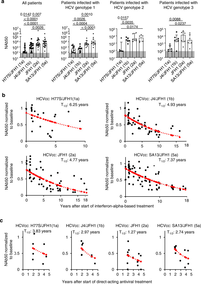
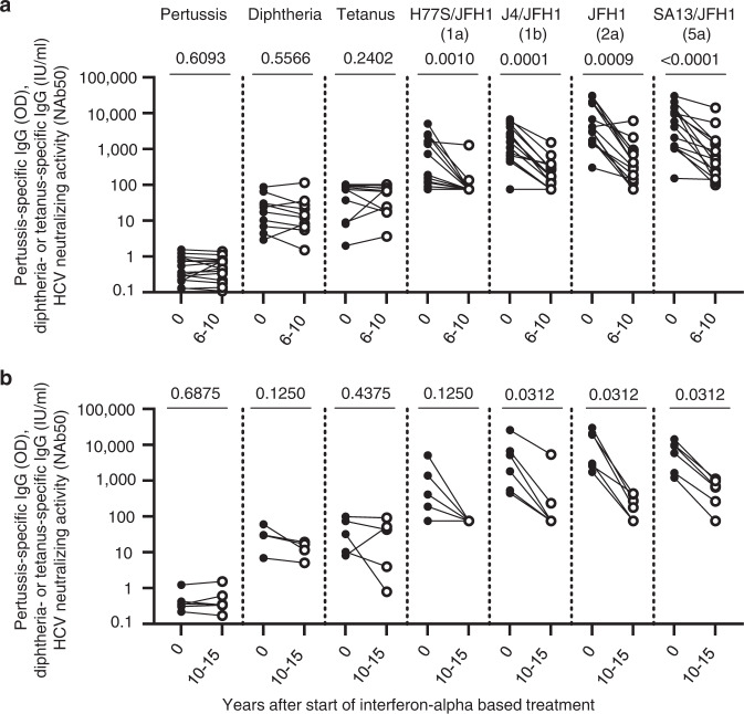
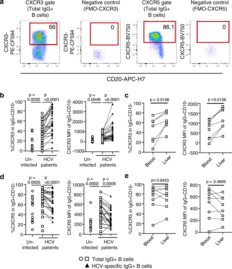
The increasing incidence of hepatitis C virus (HCV) infections underscores the need for an effective vaccine. Successful vaccines to other viruses generally depend on a long-lasting humoral response. However, data on the half-life of HCV-specific responses are lacking. Here we study archived sera and mononuclear cells that were prospectively collected up to 18 years after cure of chronic HCV infection to determine the role of HCV antigen in maintaining neutralizing antibody and B cell responses. We show that HCV-neutralizing activity decreases rapidly in potency and breadth after curative treatment. In contrast, HCV-specific memory B cells persist, and display a restored resting phenotype, normalized chemokine receptor expression and preserved ability to differentiate into antibody-secreting cells. The short half-life of HCV-neutralizing activity is consistent with a lack of long-lived plasma cells. The persistence of HCV-specific memory B cells and the reduced inflammation after cure provide an opportunity for vaccination to induce protective immunity against re-infection.


Conflict of interest statement

The authors declare no competing interests.

Figures

Fig. 1
Fig. 1. HCV-neutralizing activity declines after cure of chronic hepatitis C.
Neutralizing activity of sera from patients with chronic HCV genotype 1, 2, or 3 infections. Neutralization was tested against cell culture-produced hepatitis C viruses (HCVcc) with JFH1 backbone and core-NS2 sequences of H77S (genotype 1a), J4 (genotype 1b), JFH1 (genotype 2a), and SA13 (genotype 5a) sequence. a NAb50 (50% neutralizing activity) is shown for sera from all patients (n = 24), or stratified by chronic HCV genotype 1 (n = 12), genotype 2 (n = 6) or genotype 3 infection (n = 6). Each symbol represents a patient; error bars show median + IQR. Two-sided p values are shown and were calculated using a linear mixed model based on the log-transformation of the outcome measurement with Tukey–Kramer procedure as a post hoc test was performed. The upper line of the shaded area represents the threshold of quantitation (NAb50 = 75). b, c NAb50 of serial serum samples of patients. Neutralization was tested against HCVcc with H77S/JFH1, J4/JFH1, JFH1, and SA13/JFH1 sequences and normalized to the baseline (chronic HCV infection prior to treatment, t = 0) value. The half-life (T1/2) of NAb50 was calculated using a simple linear model of exponential decay, with the dependent variable normalized for each patient’s baseline value. Normalized NAb50 values that were predicted by mathematical modeling (see methods section) are shown as red dots. Source data are provided as a Source Data file. b Each data point represents the NAb50 of a serum sample obtained at the indicated time after interferon-alpha-based cure of chronic HCV infection. Twenty-seven (n = 27) serum samples were tested for neutralization of H77S/JFH1 HCVcc; n = 60 serum samples tested for neutralization of J4/JFH1 HCVcc; n = 68 serum samples tested for neutralization of JFH1 HCVcc and n = 71 serum samples tested for neutralization of SA13/JFH1 HCVcc. c Each data point represents the NAb50 of a serum sample obtained at the indicated time after direct-acting antiviral (DAA) therapy with n = 7 serum samples tested for neutralization of H77S/JFH1 HCVcc; n = 9 serum samples tested for neutralization of J4/JFH1 HCVcc; n = 13 serum samples tested for neutralization of JFH1 HCVcc and n = 13 serum samples tested for neutralization of SA13/JFH1 HCVcc.
Fig. 2
Fig. 2. Patients with declining HCV-neutralizing activity maintain antibodies against pertussis, diphtheria, and tetanus toxoid antigens.
a, b Humoral immune responses to pertussis, diphtheria, and tetanus toxoid antigens and HCV-neutralizing activity. Neutralization of HCVcc with JFH1 backbone and core-NS2 sequences of H77S (genotype 1a), J4 (genotype 1b), JFH1 (genotype 2a), and SA13 (genotype 5a) was assessed. Two-sided p values are shown and calculated using the Wilcoxon matched-pairs signed-rank test. OD, optical density. NAb50, 50% neutralizing activity. a Paired serum samples were studied prior to (filled circles) and 6–10 years after (open circles) start of interferon-alpha-based cure of chronic HCV infection (n = 15 sera tested against pertussis; n = 10 and n = 11 sera tested against diphtheria toxin and tetanus toxoid, respectively; n = 15 sera tested against each of the indicated HCVcc). b Paired serum samples were studied prior to (filled circles) and 10–15 years after (open circles) interferon-alpha-based cure of chronic HCV infection (n = 6 sera tested at each time point). Source data are provided as a Source Data file.
Fig. 3
Fig. 3. The breadth of serum neutralizing activity decreases after treatment-induced clearance of HCV.
a The heat map represents the neutralizing activity of serially collected sera from patients (n = 17) at 4–6 time points prior to (pre) and after the start of interferon-alpha-based treatment of chronic HCV infection. Each serum was tested against a diverse panel of 19 HCVpp listed on the y-axis. The percentage of neutralization achieved by 1:100 dilution of serum, tested in duplicate, is indicated for each HCVpp. b, c Dynamic change in the neutralization breadth at serial time points prior to (pre) and after interferon-induced HCV clearance. b Number of HCVpp that were neutralized at >25% by the sera in (a). c Average number of HCVpp neutralized with the indicated neutralization efficacy by the sera in (a). Source data are provided as a Source Data file.
Fig. 4
Fig. 4. Differential phenotype of HCV-specific and total memory B cells in HCV-infected patients and uninfected controls.
a Representative flow cytometry plots illustrating the gating strategy to identify HCV envelope 2 (E2)-specific memory B cells. b Frequency of HCV-specific B cells in the blood of patients with chronic HCV infection (n = 48) and uninfected controls (n = 17). Each data point represents one study participant; median + IQR are shown. Two-sided p values were calculated with the Mann–Whitney U-test. c Representative flow cytometry plots illustrating CD10- (mature) IgG+ (class-switched) total memory B cells and HCV-specific B cells examined for CD21 and CD27 expression. d Size of B cell subsets with a resting, intermediate, tissue-like, and activated phenotype among total or HCV-specific IgG+CD10- peripheral blood B cells of patients with chronic HCV infection (n = 48 and n = 31, respectively) and uninfected controls (n = 17). Samples with less than 30 HCV E2 tetramer+ events were excluded from CD21/CD27 subset analysis. Two-sided p values were calculated using the Wilcoxon signed-rank test was used for paired data or the Mann–Whitney U-test for unpaired data. Source data are provided as a Source Data file.
Fig. 5
Fig. 5. HCV-specific memory B cells express a liver-homing chemokine receptor profile.
a Representative flow cytometry plots illustrating CXCR3 and CXCR5 expression on IgG+ B cells. FMO, fluorescence-minus-one. b The frequencies of CXCR3+ (liver-homing) IgG+ B cells from patients with chronic HCV infection (n = 42) and uninfected controls (n = 17), and of CXCR3+ HCV-specific IgG+ B cells from patients with chronic HCV infection (n = 27) were compared (left panel). The CXCR3 geometric mean fluorescent intensity (MFI) of IgG+ B cells from patients with chronic HCV infection (n = 42) and uninfected controls (n = 17), and of HCV-specific IgG+ B cells from patients with chronic HCV infection (n = 29) were compared (right panel). c The frequencies of CXCR3+ (liver-homing) B cells (left panel) and their CXCR3 MFI (right panel) were compared in paired blood and liver specimens of patients with chronic HCV infection (n = 7 paired samples in left panel, n = 8 in right panel). d The frequencies of CXCR5+ (lymph node-homing) IgG+ B cells from patients with chronic HCV infection (n = 42) and uninfected controls (n = 17), and of CXCR5+ HCV-specific IgG+ B cells from patients with chronic HCV infection (n = 27) were compared (left panel). The CXCR5+ MFI of IgG+ B cells from patients with chronic HCV infection (n = 42) and uninfected controls (n = 17), and of HCV-specific IgG+ B cells from patients with chronic HCV infection (n = 28) were compared (right panel). e The frequencies of CXCR5+ (lymph node-homing) B cells (left panel) and their CXCR5 MFI (right panel) were compared in paired blood and liver specimens of patients with chronic HCV infection (n = 8 paired samples in each panel). Two-sided p values were calculated with the Wilcoxon signed-rank test for paired data and the Mann–Whitney U-test for unpaired data. Each data point represents one study participant. Samples with less than 30 HCV E2 tetramer+ events were excluded (be). Source data are provided as a Source Data file.
Fig. 6
Fig. 6. Phenotype of HCV-specific memory B cells after cure of chronic hepatitis C.
a, b Longitudinal change in the frequency of HCV-specific B cells prior to and after DAA- (a) or interferon-alpha-based cure of chronic hepatitis C (b). n = 10 patients were studied at 2–5 time points each (a); n = 4 patients were studied at two time points each in (b). c Differential frequency of CD21/CD27 B cell subsets within the HCV-specific and total IgG+ B cell population prior to and >3 years after HCV clearance. Paired samples from n = 8 patients were studied. d The frequency of CXCR5+ and CXCR3+ B cells and the MFI of CXCR5 or CXCR3 expression within the HCV-specific and IgG+ B cells populations prior to and >3 years after HCV clearance. Paired samples from n = 7 patients were studied. e B cells from paired PBMC samples prior to and after treatment-induced HCV clearance were differentiated into antibody-secreting cells (ASCs). The number of ASCs that secrete HCV-specific and total IgG was determined by Elispot analysis. Paired samples from n = 4 patients were studied. Two-sided p values were determined with the Wilcoxon matched-pairs signed-rank test. Source data are provided as a Source Data file.

References

    1. Bartenschlager R, et al. Critical challenges and emerging opportunities in hepatitis C virus research in an era of potent antiviral therapy: considerations for scientists and funding agencies. Virus Res. 2018;248:53–62. doi: 10.1016/j.virusres.2018.02.016. - DOI - PubMed
    1. Pawlotsky JM. Hepatitis C virus resistance to direct-acting antiviral drugs in interferon-free regimens. Gastroenterology. 2016;151:70–86. doi: 10.1053/j.gastro.2016.04.003. - DOI - PubMed
    1. World Health Organization. Guidelines for the care and treatment of persons diagnosed with chronic hepatitis C virus infection. Geneva: World Health Organization; Licence: CC BY-NC-SA 3.0 IGO. ISBN 978-92-4-155034-5 (2018). - PubMed
    1. Hill AM, Nath S, Simmons B. The road to elimination of hepatitis C: analysis of cures versus new infections in 91 countries. J. Virus Erad. 2017;3:117–123. doi: 10.1016/S2055-6640(20)30329-0. - DOI - PMC - PubMed
    1. Falade-Nwulia O, Sulkowski M. The HCV care continuum does not end with cure: A call to arms for the prevention of reinfection. J. Hepatol. 2017;66:267–269. doi: 10.1016/j.jhep.2016.10.027. - DOI - PMC - PubMed

Publication types

Substances