. 2022 Sep 16;13(1):5451.
doi: 10.1038/s41467-022-32951-4.

Structure-based discovery of small molecules that disaggregate Alzheimer's disease tissue derived tau fibrils in vitro

Affiliations

Structure-based discovery of small molecules that disaggregate Alzheimer's disease tissue derived tau fibrils in vitro

Paul M Seidler et al. Nat Commun. .

Abstract

Alzheimer's disease (AD) is the consequence of neuronal death and brain atrophy associated with the aggregation of protein tau into fibrils. Thus disaggregation of tau fibrils could be a therapeutic approach to AD. The small molecule EGCG, abundant in green tea, has long been known to disaggregate tau and other amyloid fibrils, but EGCG has poor drug-like properties, failing to fully penetrate the brain. Here we have cryogenically trapped an intermediate of brain-extracted tau fibrils on the kinetic pathway to EGCG-induced disaggregation and have determined its cryoEM structure. The structure reveals that EGCG molecules stack in polar clefts between the paired helical protofilaments that pathologically define AD. Treating the EGCG binding position as a pharmacophore, we computationally screened thousands of drug-like compounds for compatibility for the pharmacophore, discovering several that experimentally disaggregate brain-derived tau fibrils in vitro. This work suggests the potential of structure-based, small-molecule drug discovery for amyloid diseases.


Conflict of interest statement

D.S.E. is SAB chair and equity holder of ADRx, Inc. All other authors declare no conflicts.

Figures
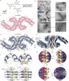
Fig. 1
Fig. 1. CryoEM structure of AD-tau PHF in complex with EGCG.
a Epigallocatechin gallate (EGCG) is the most abundant polyphenol in green tea. It includes a benzenediol ring (A) adjoined to a tetrahydropyran moiety (C), which are connected to a galloyl ring (D) and pyrogallol ring (B). The 8 hydroxyl groups allow EGCG to engage in hydrogen bonding and other polar interactions with numerous biomolecules. b Electron micrographs of brain-derived PHFs over the course of EGCG incubation. Without EGCG (top), numerous fibrils are observed (representative image from n = 15). After 3-h incubation at 37  °C (middle), subtle changes in the fibril morphology are present, with a widening of the fibril fuzzy coat (representative image from n = 15). Far fewer fibrils are seen at this time point. After overnight EGCG incubation (bottom), the rare remaining fibrils appear swollen and disturbed (representative image from n = 8). c Cross-sectional view of the AD patient brain-derived tau PHF cryoEM structure before the addition of EGCG. d Tau PHF structure following 3-h incubation with EGCG. Three new regions of density become apparent with the addition of EGCG (Sites 1–3). Site 1 is located in the polar cleft at the intersection of the two protofilaments composing the PHF. Sites 2 and 3 of new density are observed adjacent to K343 and K347 near the β-helix of the fibril. Both Sites 2 and 3 display weaker density than Site 1. e Tilted view of the 3-h structure with EGCG bound at Site 1. f Close up top- and side-views of EGCG in Site 1. This region borders N327, H329, E338, and K340 of the fibril, with EGCG making polar and hydrogen-bond contacts with these residues. EGCG adopts a primarily planar conformation when bound to the fibril, stabilized by π-π interactions of the stacked aromatic rings of EGCG. When viewed from the side of the fibril axis, the EGCG density is seen stacking with the same period as the fibril layers. g Side-view of a single EGCG molecule buried by the fibril and other copies of EGCG.
Fig. 2
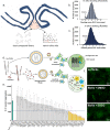
Fig. 2. In silico and in vitro screening of tau disaggregants using EGCG pharmacophore.
a To identify novel compounds capable of fibril disaggregation, we performed an in silico screen using the EGCG binding site to the tau PHF (red circle). Two libraries of compounds were docked to the site using two computational methods (AutoDock and Rosetta), and hits were ranked and selected for experimental characterization. b, c Distribution of in silico docking scores of compound libraries using AutoDock (b) and Rosetta (c). For both methods, more negative scores indicate stronger compound binding. EGCG was a control for each method. As shown, both methods identify EGCG as a strong binder to the site on the tau PHF. d Top hits from the computational screen were selected for experimental characterization. Compounds were initially screened using an in vitro biosensor cell assay. Brain-derived tau fibrils were incubated with and without an inhibitor compound. Fibrils were then dissolved in liposomes and transduced into HEK293T cells overexpressing fluorescently labeled tau. When fibrils are transfected into the cells, the exogenous seeds initiate the aggregation of the endogenous tau, resulting in the formation of intracellular fluorescent puncta. If fibrils are effectively disaggregated by an inhibitor compound, the exogenous fibril seeds will be dissolved, and the intracellular tau will remain soluble, with no puncta formed. e Quantification of hit compounds in tau biosensor cell assay. For fibrils treated with DMSO vehicle control (turquoise bar), many fluorescent aggregates are seen. Without the addition of fibrils, (“no seed”), no intracellular aggregation occurs. Incubation of fibrils with EGCG also prevents the formation of any seeds. The dashed line indicates a 50% reduction in the number of aggregates. Yellow bars indicate any compound that produces a >50% reduction in aggregate formation. Error bars represent ±SD, all experiments were performed with n = 3 experimental replicates. f Fluorescent microscopy images of biosensor cells without fibril seeds added (top), with seeds and DMSO control (middle), and with seeds and EGCG (bottom). Numerous bright intracellular puncta are seen in the DMSO control, which are eliminated with the addition of EGCG.
Fig. 3
Fig. 3. Characterization of tau disaggregation by lead compounds.
a Top hits from the in silico and biosensor cell screens were selected for further experimental characterization. Four compounds were selected, CNS-11, CNS-17, CNS-2, and CNS-12. b Electron micrographs of brain-derived tau fibrils after incubation with each compound, with EGCG as control. Few fibrils are observed with EGCG treatment, as well as with CNS-11 and CNS-12. Scale bars represent 250 nm. c Quantitation of fibril number present on EM images with and without compound treatment. N = 33 images were taken from random points on the EM grid, and fibrils were counted. Error bars represent the standard deviation of triplicate technical measurements. A large reduction in visible fibrils is seen for CNS-11. d Brain-derived tau fibrils were treated with compound and the insoluble fraction was analyzed by Western blot, staining for total tau. e Quantitation of insoluble tau abundance in the Western blot. Similarly, both EGCG and the lead compounds substantially reduce the amount of insoluble tau in the fraction. N = 3 experimental replicates were performed for each treatment condition. f MTT cytotoxicity assay in Neuro2a cell model. Brain-derived tau PHFs with and without vehicle control (PBS) show no toxicity (blue bars). Compounds alone show varied toxicities (dark orange bars). Compounds incubated with tau fibrils (light orange bars) do not show additional toxicity. All error bars represent ±SD. N = 3 experimental replicates were performed for each g Model of CNS-11 docked to the EGCG binding site on the tau PHF. CNS-11 is within the hydrogen bonding distance of both H329 and K340. For c, e, f *p < 0.05, **p < 0.01, ***p < 0.001 using a one-way ANOVA.
Fig. 4
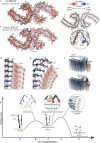
Fig. 4. Structure-informed mechanism for EGCG-driven disaggregation of AD-tau PHF.
Solvation energy calculations of tau PHF structures without EGCG (a) and with EGCG after 3-h incubation (b). Red residues are more stable; blue residues are less stable. The most stable residues seen across both structures are hydrophobic and buried within the fibril core, and less stable residues are typically on the solvent-exposed surface. At 3-h incubation, the structure is less stable (−28.1 kcal/mol/chain) than without EGCG (−34.9 kcal/mol/chain). c To understand the localized effects in fibril stability, energy difference maps were calculated. Subtraction of the no-EGCG model from the 3-h EGCG model shows a large increase in free energy of Lys340 at the EGCG binding site, indicating the presence of EGCG significantly destabilizes Lys340. d The 3-h PHF-EGCG structure reveals EGCG molecules stacked 4.8 Å apart, permitting each EGCG molecule to H-bond with individual stacked molecules of tau (dashed yellow lines connecting EGCG to tau side chains Asn327 and His329). The 4.8 Å spacing between tau molecules is characteristic of the intermolecular β-sheet hydrogen bonding distance (dashed yellow lines connecting tau molecules). However, this 4.8 Å spacing incurs unfavorable voids between EGCG rings A, C, and D (as indicated by gaps between space-filling atoms). e The voids between stacked aromatic groups can be filled by compressing the distance between these A, C, and D aromatic rings that face the solvent. In so doing, the EGCG stack curves, widening the spacing on the fibril-facing surface. Asn327 and His329 can maintain favorable hydrogen bonding with the curved stack of EGCG molecules only if the tau molecules separate wider than 4.8 Å. This separation would allow water to solvate the separated tau molecules. The curvature of the EGCG stack fills the unfavorable voids between EGCG aromatic rings and further widens the separation between tau molecules. By this mechanism, the binding energy between stacked EGCG molecules is converted to a conformational change that pries apart stacked tau molecules. f Alternate view showing a tau PHF protofilament being disrupted by the curvature of stacked EGCG molecules. g Reaction coordinate diagram describing the possible mechanism of tau disaggregation by EGCG. Tau PHFs in solution with EGCG (coordinate A) are bound by repeating stacks of EGCG molecules (coordinate B). Once EGCG is bound, local charge-mediated effects begin to destabilize the fibril (coordinate C). These effects include unfavorable burying of charged residues (e.g. Lys340) and disruption of pairing between charged side chains. These repulsive forces, in addition to possible backbone H-bonding between tau and EGCG, weaken the β-sheet H-bond network of the fibril. Lastly, conformational changes induced by EGCG π-π stacking (described in df) may further disrupt the fibril architecture (coordinate D), leading to the disaggregated EGCG-bound tau end product (coordinate E).

References

    1. La Joie R, et al. Prospective longitudinal atrophy in Alzheimer’s disease correlates with the intensity and topography of baseline tau-PET. Sci. Transl. Med. 2020;12:eaau5732. doi: 10.1126/scitranslmed.aau5732. - DOI - PMC - PubMed
    1. Chang C-W, Shao E, Mucke L. Tau: enabler of diverse brain disorders and target of rapidly evolving therapeutic strategies. Science. 2021;371:eabb8255. doi: 10.1126/science.abb8255. - DOI - PMC - PubMed
    1. Soeda, Y. & Takashima, A. New insights into drug discovery targeting tau protein. Front. Mol. Neurosci. 13, 590896 (2020). - PMC - PubMed
    1. Congdon EE, Sigurdsson EM. Tau-targeting therapies for Alzheimer disease. Nat. Rev. Neurol. 2018;14:399–415. doi: 10.1038/s41582-018-0013-z. - DOI - PMC - PubMed
    1. LMTM. ALZFORUM. https://www.alzforum.org/therapeutics/lmtm (2019).

Publication types