. 2022 Sep 16;13(1):5449.
doi: 10.1038/s41467-022-33227-7.

Molecular architecture of the augmin complex

Affiliations

Molecular architecture of the augmin complex

Clinton A Gabel et al. Nat Commun. .

Abstract

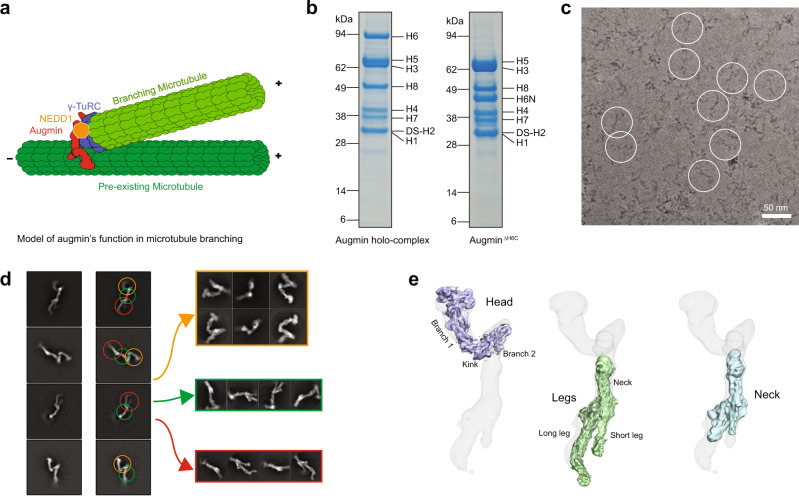
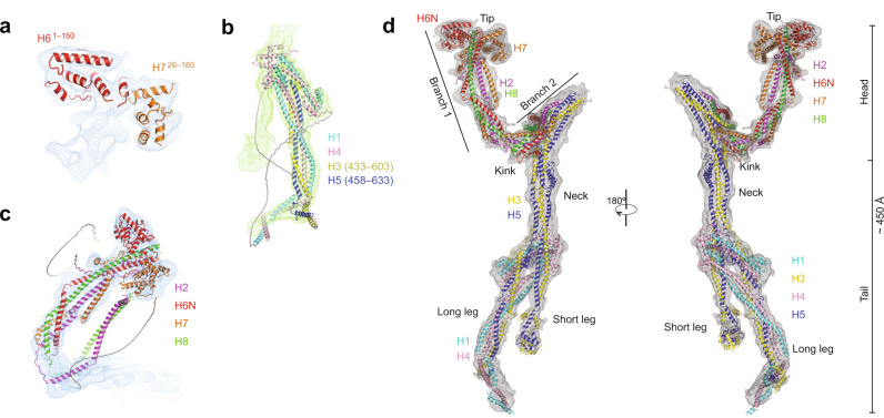
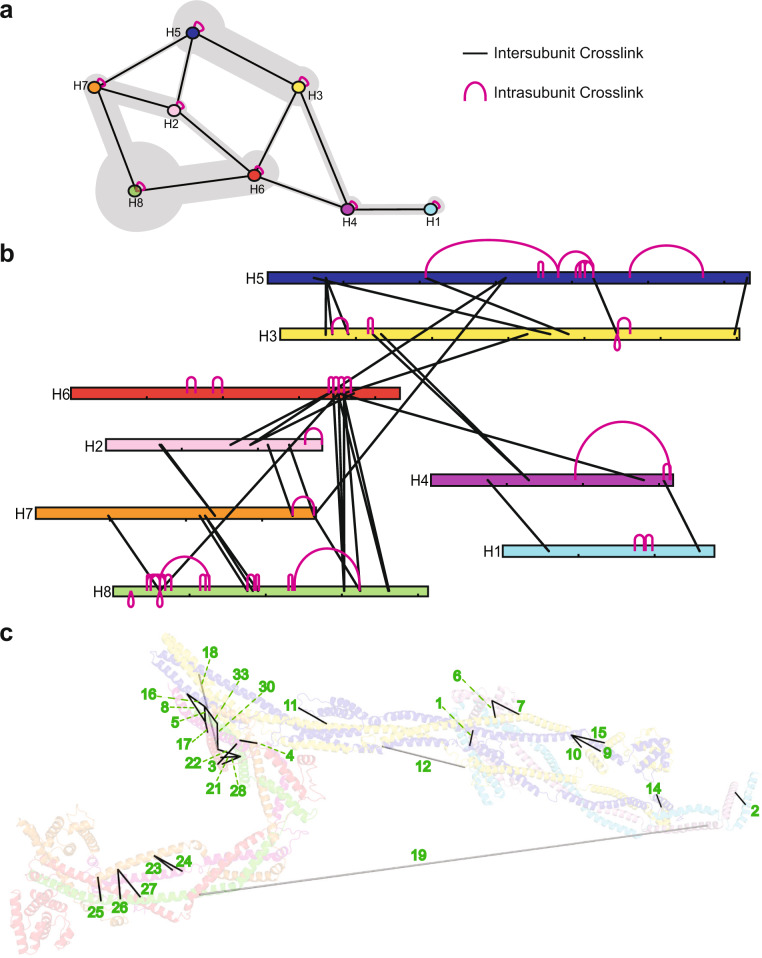
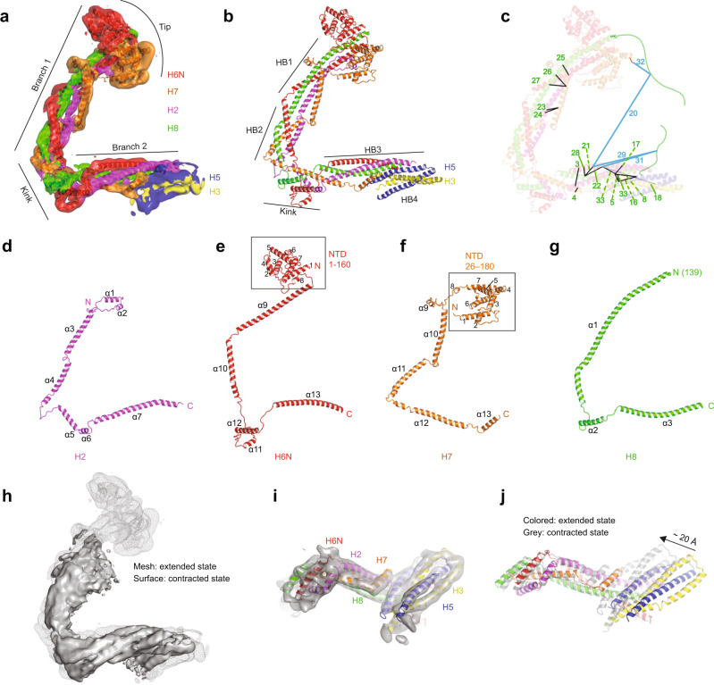
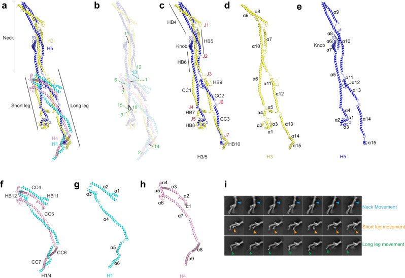
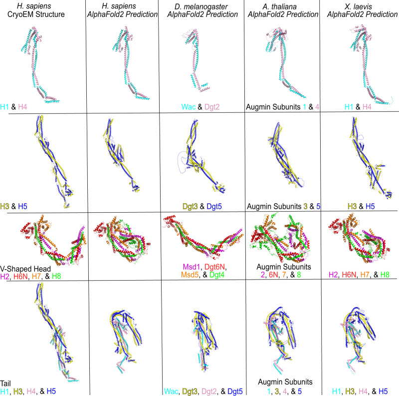
Accurate segregation of chromosomes during mitosis depends on the correct assembly of the mitotic spindle, a bipolar structure composed mainly of microtubules. The augmin complex, or homologous to augmin subunits (HAUS) complex, is an eight-subunit protein complex required for building robust mitotic spindles in metazoa. Augmin increases microtubule density within the spindle by recruiting the γ-tubulin ring complex (γ-TuRC) to pre-existing microtubules and nucleating branching microtubules. Here, we elucidate the molecular architecture of augmin by single particle cryo-electron microscopy (cryo-EM), computational methods, and crosslinking mass spectrometry (CLMS). Augmin's highly flexible structure contains a V-shaped head and a filamentous tail, with the head existing in either extended or contracted conformational states. Our work highlights how cryo-EM, complemented by computational advances and CLMS, can elucidate the structure of a challenging protein complex and provides insights into the function of augmin in mediating microtubule branching nucleation.


Conflict of interest statement

The authors declare no competing interests.

Figures

Fig. 1
Fig. 1. Purification and cryo-EM reconstruction of augmin.
a A diagram of augmin–γ-TuRC–NEDD1 binding to pre-existing microtubules to create shallow-angled branching microtubules. b Representative SDS–PAGE gels of augmin holo-complex and augminΔH6C. The results shown are representative of more than three experiments. The uncropped gel is provided in the Source Data file. c Representative cryo-EM micrograph of augminΔH6C from a total of 20,021 micrographs. Scale bar 50 nm. Examples of augmin particles are circled. d 2D classification of augminΔH6C where classes are segmented into V-shaped head (orange arrow), neck of the tail (green arrow), and legs of the tail (red arrow) before further 3D classification and refinement. e Cryo-EM maps of segmented regions including the V-shaped head (light blue), the legs (green), and the neck (cyan), superimposed to a low-resolution reconstruction of the whole complex (gray mesh).
Fig. 2
Fig. 2. Fitting of HAUS subunits and full atomic model of augmin.
a Rigid-body fitting of the individual AlphaFold2 predictions of H6 (a.a. 1–160) and H7 (a.a. 26–160) within the cryo-EM map of the tip of branch 1. b Rigid-body fitting of the ColabFold prediction of the long leg. c Rigid-body fitting of the ColabFold prediction of branch 1 of the V-shaped head. d Atomic model of augmin superimposed to a combined cryo-EM map (in gray mesh).
Fig. 3
Fig. 3. Crosslinking mass spectrometry of augminΔH6C.
a Summary of crosslinks between HAUS subunits. The thickness of gray shading indicates the number of crosslinks. b Relative positions of each individual intrasubunit (pink) and intersubunit (black) crosslink identified by CLMS. c Intersubunit crosslinks are mapped onto the augmin structure using PyXlinkViewer. Subunits are colored as in panels (a), (b). Labels correspond to numbers in Table 1. Black lines represent crosslink Cα–Cα distances <30 Å and light gray lines represent distances >30 Å.
Fig. 4
Fig. 4. Structure of the V-shaped head of augmin.
a Structure of the V-shaped head in extended conformation superimposed into the cryo-EM map with each subunit color-coded. b Structure of the V-shaped head in extended conformation in cartoon presentation. c Intersubunit crosslinks as labeled in Table 1 are mapped on the structure. Intersubunit crosslinks below 30 Å (black), above 30 Å (light gray), and those with one peptide from the N- and C-termini of H8 (drawn as green disordered chains) that are not modeled within augmin’s structure (blue). d H2 with labeled helices numbered from N to C-terminus (α1–7). e H6N with labeled α-helices numbered from N to C-terminus (α1–13). NTD (a.a. 1–160, α1–8) enclosed within a box. f H7 with labeled α-helices numbered from N to C-terminus (α1–13). NTD (a.a. 1–160, α1–8) enclosed within a box. g H8 with labeled α-helices numbered from N to C-terminus (α1–3). h Overlayed cryo-EM maps of the extended state (mesh, gray) and contracted state (surface, gray). i Bottom view of the V-shaped head in the contracted state. j Structural comparison of the V-shaped head in the extended and contracted state. A conformational shift of ~20 Å in the contracted state (gray) compared to the extended state (colored) is observed.
Fig. 5
Fig. 5. Structure of the tail of augmin.
a Overall architecture of the tail of augmin with helices from H1/3/4/5 depicting the neck, short, and long legs. b Intersubunit crosslinks as labeled in Table 1 are mapped on the structure. Intersubunit crosslinks below 30 Å (black), above 30 Å (light gray), and one containing a peptide from a H5 region not modeled within augmin’s structure (blue). c H3/5 dimer with labeled features: CC1–3, HB4–8, J1–7, and the knob. d H3 with labeled α-helices numbered from N to C-terminus (α1–15). e H5 with labeled α-helices numbered from N to C-terminus (α1–15). f H1/4 dimer with labeled features: CC4–7 and HB11–12. g H1 with labeled α-helices numbered from N to C-terminus (α1–6). h H4 with labeled α-helices numbered from N to C-terminus (α1–9). i 2D classes depicting independent movement of the neck and the short and long legs.
Fig. 6
Fig. 6. Cross-species comparisons of the augmin complex predicted by AlphaFold2.
Comparisons of the H1/4 and H3/5 dimers, V-shaped head, and tail of augmin across H. sapiens, D. melanogaster, A. thaliana, and X. laevis. Cryo-EM structures of the H. sapiens H1/4 and H3/5 dimers and V-shaped head and tail regions are provided as references.

References

    1. Petry S. Mechanisms of mitotic spindle assembly. Annu. Rev. Biochem. 2016;85:659–683. doi: 10.1146/annurev-biochem-060815-014528. - DOI - PMC - PubMed
    1. Goshima G, et al. Genes required for mitotic spindle assembly in Drosophila S2 cells. Science. 2007;316:417–421. doi: 10.1126/science.1141314. - DOI - PMC - PubMed
    1. Goshima G, et al. The augmin complex plays a critical role in spindle microtubule generation for mitotic progression and cytokinesis in human cells. Proc. Natl Acad. Sci. USA. 2009;106:6998–7003. doi: 10.1073/pnas.0901587106. - DOI - PMC - PubMed
    1. Petry S, Groen AC, Ishihara K, Mitchison TJ, Vale RD. Branching microtubule nucleation in Xenopus egg extracts mediated by augmin and TPX2. Cell. 2013;152:768–777. doi: 10.1016/j.cell.2012.12.044. - DOI - PMC - PubMed
    1. Petry S, Pugieux C, Nedelec FJ, Vale RD. Augmin promotes meiotic spindle formation and bipolarity in Xenopus egg extracts. Proc. Natl Acad. Sci. USA. 2011;108:14473–14478. doi: 10.1073/pnas.1110412108. - DOI - PMC - PubMed

Publication types

MeSH terms