. 2022 Sep 16;12(1):15550.
doi: 10.1038/s41598-022-19464-2.

Optimal flickering light stimulation for entraining gamma rhythms in older adults

Affiliations

Optimal flickering light stimulation for entraining gamma rhythms in older adults

Yeseung Park et al. Sci Rep. .

Abstract

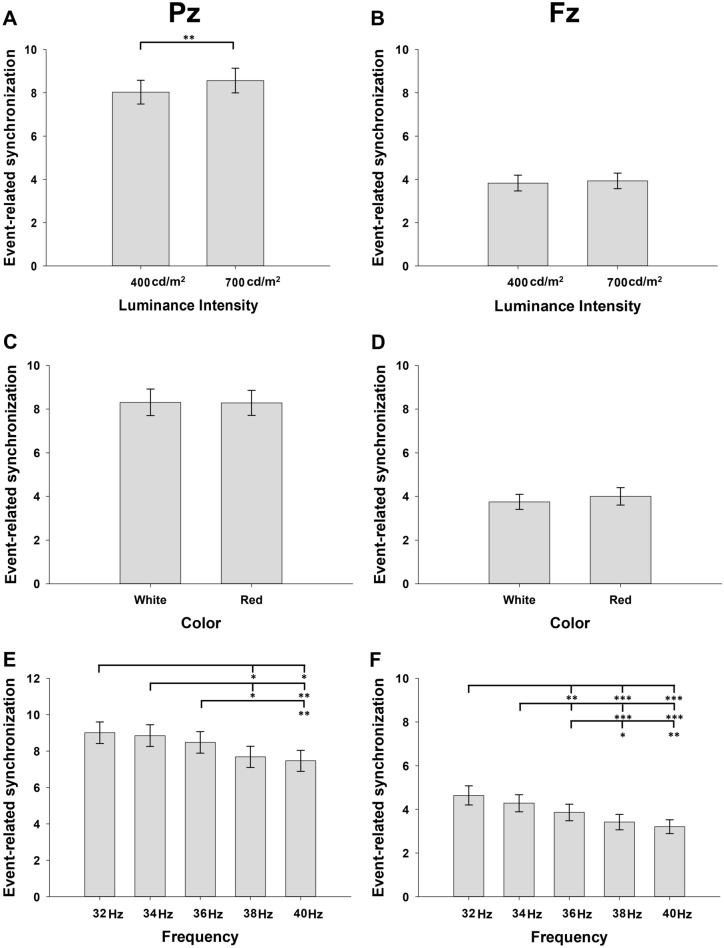
With aging, optimal parameters of flickering light stimulation (FLS) for gamma entrainment may change in the eyes and brain. We investigated the optimal FLS parameters for gamma entrainment in 35 cognitively normal old adults by comparing event-related synchronization (ERS) and spectral Granger causality (sGC) of entrained gamma rhythms between different luminance intensities, colors, and flickering frequencies of FLSs. ERS entrained by 700 cd/m2 FLS and 32 Hz or 34 Hz FLSs was stronger than that entrained by 400 cd/m2 at Pz (p < 0.01) and 38 Hz or 40 Hz FLSs, respectively, at both Pz (p < 0.05) and Fz (p < 0.01). Parieto-occipital-to-frontotemporal connectivities of gamma rhythm entrained by 700 cd/m2 FLS and 32 Hz or 34 Hz FLSs were also stronger than those entrained by 400 cd/m2 at Pz (p < 0.01) and 38 Hz or 40 Hz FLSs, respectively (p < 0.001). ERS and parieto-occipital-to-frontotemporal connectivities of entrained gamma rhythms did not show significant difference between white and red lights. Adverse effects were comparable between different parameters. In older adults, 700 cd/m2 FLS at 32 Hz or 34 Hz can entrain a strong gamma rhythm in the whole brain with tolerable adverse effects.


Conflict of interest statement

The authors declare no competing interests.

Figures

Figure 1
Figure 1
Comparison of grand-average event-related spectral perturbation (ERSP) of steady-state visually evoked potentials induced by flickering light stimulation (FLS) between colors, luminance intensities, and flickering frequencies. Each column shows ERSP from 750 ms before the onset of FLS to 750 ms after the offset of FLS.
Figure 2
Figure 2
Main effect of time windows on grand-average event-related synchronization (ERS) of steady-state visually evoked potentials induced by flickering light stimulation (FLS). T0–T10 indicates 250 ms time windows from 250 ms before the onset of FLS to 500 ms after the offset of FLS.
Figure 3
Figure 3
Main effects of the luminance intensities, colors, and flickering frequencies of flickering light stimulations on event-related synchronization (ERS) (A) and (B) demonstrate the main effect of luminance intensities, (C) and (D) demonstrate the main effect of colors, (E) and (F) demonstrate the main effect of flickering frequencies. Error bars indicate standard errors. *p < 0.05, **p < 0.01, and ***p < 0.001 by repeated-measures analysis of variance.
Figure 4
Figure 4
Changes in the gamma rhythm connectivity after flickering light stimulation. The spectral Granger causality (sGC) is analyzed in 1953 connections between 63 electrodes. The numbers electrodes from 1 to 63 correspond to Fp1, Fp2, AF7, AF3, AFz, AF4, AF8, F7, F5, F3, F1, Fz, F2, F4, F6, F8, FT9, FT7, FC5, FC3, FC1, FC2, FC4, FC6, FT8, FT10, T7, C5, C3, C1, Cz, C2, C4, C6, T8, TP9, TP7, CP5, CP3, CP1, CPz, CP2, CP4, CP6, TP8, TP10, P7, P5, P3, P1, P2, P4, P6, P8, PO7, PO3, POz, PO4, PO8, O1, Oz, and O2, respectively. (A) t-values in the spectral Granger causality of the occipitoparietal to frontotemporal gamma rhythm connections. The left-upper side of each matrix represents the occipitoparietal to frontotemporal connections. (B) Main effect of luminance intensity on the averaged strength of the occipitoparietal to frontotemporal gamma rhythm connections; (C) Main effect of flickering frequency on the averaged strength of the occipitoparietal to frontotemporal gamma rhythm connections. Error bars indicate standard errors. ***p < 0.001 by repeated-measures analysis of variance.
Figure 5
Figure 5
(A) Experimental procedures FLS, flickering light stimulation; rsEEG, resting-state electroencephalogram; BR, break (B) An example of a subject wearing a flickering light stimulation device.

References

    1. Rodriguez E, et al. Perception’s shadow: Long-distance synchronization of human brain activity. Nature. 1999;397:430–433. doi: 10.1038/17120. - DOI - PubMed
    1. Baldauf D, Desimone R. Neural mechanisms of object-based attention. Science. 2014;344:424–427. doi: 10.1126/science.1247003. - DOI - PubMed
    1. Kucewicz MT, et al. Dissecting gamma frequency activity during human memory processing. Brain. 2017;140:1337–1350. doi: 10.1093/brain/awx043. - DOI - PubMed
    1. Roux F, Wibral M, Mohr HM, Singer W, Uhlhaas PJ. Gamma-band activity in human prefrontal cortex codes for the number of relevant items maintained in working memory. J. Neurosci. 2012;32:12411–12420. doi: 10.1523/jneurosci.0421-12.2012. - DOI - PMC - PubMed
    1. Schneider TR, Debener S, Oostenveld R, Engel AK. Enhanced EEG gamma-band activity reflects multisensory semantic matching in visual-to-auditory object priming. Neuroimage. 2008;42:1244–1254. doi: 10.1016/j.neuroimage.2008.05.033. - DOI - PubMed

Publication types