

Prediction of visual function from automatically quantified optical coherence tomography biomarkers in patients with geographic atrophy using machine learning
- PMID: 36114218
- PMCID: PMC9481631
- DOI: 10.1038/s41598-022-19413-z
Prediction of visual function from automatically quantified optical coherence tomography biomarkers in patients with geographic atrophy using machine learning
Abstract
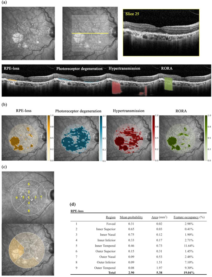
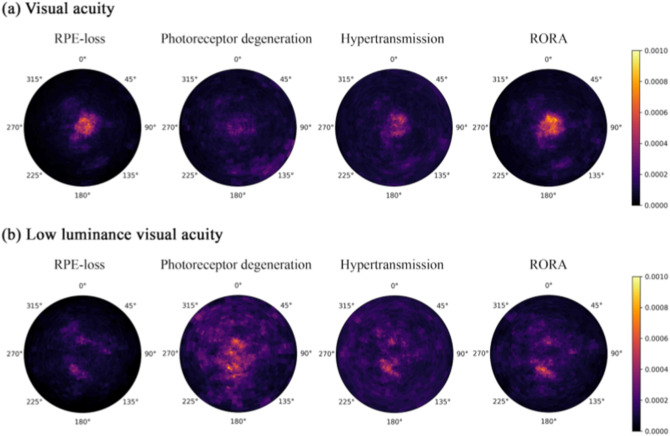
Geographic atrophy (GA) is a vision-threatening manifestation of age-related macular degeneration (AMD), one of the leading causes of blindness globally. Objective, rapid, reliable, and scalable quantification of GA from optical coherence tomography (OCT) retinal scans is necessary for disease monitoring, prognostic research, and clinical endpoints for therapy development. Such automatically quantified biomarkers on OCT are likely to further elucidate structure-function correlation in GA and thus the pathophysiological mechanisms of disease development and progression. In this work, we aimed to predict visual function with machine-learning applied to automatically acquired quantitative imaging biomarkers in GA. A post-hoc analysis of data from a clinical trial and routine clinical care was conducted. A deep-learning automated segmentation model was applied on OCT scans from 476 eyes (325 patients) with GA. A separate machine learning prediction model (Random Forest) used the resultant quantitative OCT (qOCT) biomarkers to predict cross-sectional visual acuity under standard (VA) and low luminance (LLVA). The primary outcome was regression coefficient (r2) and mean absolute error (MAE) for cross-sectional VA and LLVA in Early Treatment Diabetic Retinopathy Study (ETDRS) letters. OCT parameters were predictive of VA (r2 0.40 MAE 11.7 ETDRS letters) and LLVA (r2 0.25 MAE 12.1). Normalised random forest feature importance, as a measure of the predictive value of the three constituent features of GA; retinal pigment epithelium (RPE)-loss, photoreceptor degeneration (PDR), hypertransmission and their locations, was reported both on voxel-level heatmaps and ETDRS-grid subfields. The foveal region (46.5%) and RPE-loss (31.1%) had greatest predictive importance for VA. For LLVA, however, non-foveal regions (74.5%) and PDR (38.9%) were most important. In conclusion, automated qOCT biomarkers demonstrate predictive significance for VA and LLVA in GA. LLVA is itself predictive of GA progression, implying that the predictive qOCT biomarkers provided by our model are also prognostic.
© 2022. The Author(s).
Conflict of interest statement
NP: Moorfields Eye Charity Career Development Award (R190031A), equity owner, Phenopolis Ltd. DJF: Consulting for Abbvie, Allergan, DeepMind. LF: Nothing to declare. GZ: No conflicts of interest. BL: No conflicts of interest. SG: Moorfields Eye Charity Grant (GR001003), Wellcome Trust Grant (206619_Z_17_Z). SKW: MRC Clinical Research Training Fellowship (MR/T000953/1). RS: No conflicts of interest. PAK: Moorfields Eye Charity Career Development Award (R190028A), UK Research & Innovation Future Leaders Fellowship (MR/T019050/1); Consulting for DeepMind, Roche, Novartis, Apellis and BitFount; equity owner in Big Picture Medical; speaker fees from Heidelberg Engineering, Topcon, Allergan, and Bayer. KB: Speaker fees from Novartis, Bayer, Alimera, Allergan and Heidelberg, consulting Novartis and Roche and research support from Apellis, Novartis and Bayer. PJP: Speaker fees from Bayer, Heidelberg, Roche and Topcon, consulting Bayer, Novartis, Oxford Bioelectronics and Roche and research support from Bayer. AM: Employee of Apellis. TK: No conflicts of interest.
Figures
References
Publication types
MeSH terms
Substances
Grants and funding
LinkOut - more resources
Full Text Sources
