. 2023 Jan;30(1):184-194.
doi: 10.1038/s41418-022-01057-1. Epub 2022 Sep 16.

Bud31-mediated alternative splicing is required for spermatogonial stem cell self-renewal and differentiation

Affiliations

Bud31-mediated alternative splicing is required for spermatogonial stem cell self-renewal and differentiation

Junchao Qin et al. Cell Death Differ. 2023 Jan.

Abstract

Alternative splicing (AS) is tightly regulated during cell differentiation and development. AS events are prevalent in the testis, but the splicing regulation in spermatogenesis remains unclear. Here we report that the spliceosome component Bud31 plays a crucial role during spermatogenesis in mice. Germ cell-specific knockout of Bud31 led to loss of spermatogonia and to male infertility. We further demonstrate that Bud31 is required for both spermatogonial stem cell pool maintenance and the initiation of spermatogenesis. SMART-seq revealed that deletion of Bud31 in germ cells causes widespread exon-skipping and intron retention. Particularly, we identified Cdk2 as one of the direct splicing targets of Bud31, knockout of Bud31 resulted in retention of the first intron of Cdk2, which led to a decrease in Cdk2 expression. Our findings suggest that Bud31-mediated AS within spermatogonial stem cells regulates the self-renewal and differentiation of male germ cells in mammals.


Conflict of interest statement

The authors declare no competing interests.

Figures
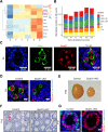
Fig. 1
Fig. 1. Germ cell-specific Bud31 knockout results in complete male infertility.
A Heatmap analysis of the expression of known splicing factors at E18 and at different postnatal day points (P0, P3, P14, P28, and P63) in mouse testes using the Evo-devo mammalian organs database (https://apps.kaessmannlab.org/evodevoapp/). B Histogram showing the occurrence of AS events at E18.5, P0, P3, P14, P28, and P63 in mouse testes using the Evo-devo mammalian organs database. C Immunofluorescence staining of Bud31 (red) and the germ cell marker Dazl (green) in the seminiferous tubules of P4 mice. The scale bar is 20 μm. D Immunofluorescence staining of Bud31 (red) and the germ cell marker Dazl (green) in seminiferous tubules of P4 testes of Bud31-vKO mice and in littermate controls. The scale bar is 10 μm. E Testes from P46 Bud31-vKO mice compared to their respective littermate controls. F Hematoxylin staining of the seminiferous tubules of the testes of P12 Bud31-vKO and littermate control mice. The scale bar is 20 μm. Red arrow: germ cells. G Immunofluorescence staining of the Sertoli cell marker Sox9 (red) and germ cell nuclear antigen Gcna (green) in the seminiferous tubules of P10 Bud31-vKO and their littermate controls. The scale bar is 10 μm.
Fig. 2
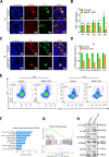
Fig. 2. Bud31 knockout leads to severe defects in SSC self-renewal.
A Immunofluorescence staining of Bud31 (red) and Plzf (green) in seminiferous tubules of P4 mice. The scale bar is 10 μm. B Quantification of Plzf-positive cells in seminiferous tubules from P1 to P5 mice assessed by immunofluorescence staining. Counts shown are Plzf-positive cells per tubule. At least 60 tubules were counted from at least three different mice. Student’s t test, error bars indicate the SEM. ***P < 0.001. C Immunofluorescence staining of SSC markers Plzf (red) and Lin28a (green) in seminiferous tubules of P5 mice. The scale bar is 10 μm. D qPCR to measure the expression of SSC self-renewal-related genes (Plzf, Lin28a, Gfrα1, and Id4) in P4 Bud31-vKO mice compared to littermate controls. Student’s t tests were performed, and data are shown as the mean ± SEM of three independent experiments. *P < 0.05, **P < 0.01, ***P < 0.001. E The spermatogonial stem cell proportion was analyzed by assessing cells positive for the SSC marker Thy1 and negative for the differentiated spermatogonia marker c-Kit using flow cytometry in P3 and P4 Bud31-vKO mice (compared against littermate controls). F Gene Ontology analysis of differentially expressed genes (DEGs) in P4 mouse testes from SMART-seq analysis of SSCs. G Gene Set Enrichment Analysis of P13K-AKT-mTOR enrichment in P4 mouse testes from the SMART-seq dataset. H Western blotting analysis of mTOR pathway-related protein expression in testes from Bud31-vKO mice. Gapdh was used as the loading control.
Fig. 3
Fig. 3. Bud31-deficient male germ cells fail to enter meiosis.
A Hematoxylin staining of the seminiferous tubules of the testes of P10 Bud31-sKO and littermate control mice. The scale bar is 50 μm. B Immunofluorescence staining of the meiotic marker γH2AX (green) and syndicate complex marker SYCP3 (red) in P12 testis tissue sections. The scale bar is 10 μm. C Immunofluorescence staining of SYCP3 (red) and γH2AX (green) was performed on chromosome spreads of Bud31-sKO mice. The scale bar is 5 μm. D Immunofluorescence staining of Stra8 (green) in the testicular tissue sections of P10 control and Bud31-sKO mice. The scale bar is 20 μm. E The expression of Stra8 and its downstream targets was assessed by qPCR in the testes of P10 Bud31-sKO and littermate control mice. The Stra8 expression by qPCR (F) and western blotting (G) in P4 testes of Bud31-vKO mice and littermate controls. *P < 0.05; **P < 0.01; ***P < 0.001.
Fig. 4
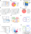
Fig. 4. Bud31 regulates AS in spermatogenic cells.
A Schematic diagram of AS types. B Pie chart depicting the proportions of different types of AS events in the SMART-seq data from spermatogenic cells in the testes of P4 Bud31-vKO mice and littermate controls. C Mapping reads coverage of the SMART-seq data in spermatogenic cells in the testes of P4 Bud31-vKO mice and littermate controls. The percentages of reads aligned to intergenic, intron, and exon regions are shown. D Global splicing efficiency at the 5′ splicing sites and 3′ splicing sites was analyzed with splicing efficiency analysis and annotation in spermatogenic cells in the testes of P4 Bud31-vKO mice and littermate controls. E Pie chart of the distribution of Bud31 binding peaks from RIP-seq data for primary cells in P14 wild-type mouse testes. F The normalized reads intensity distribution around the 3′ and 5′ splicing sites (±100 bp) was analyzed using RIP-seq data in primary cells of P14 wild-type mouse testes. G HOMER de novo motif analysis of Bud31 binding peaks based on the RIP-seq data. Two significant motifs with their corresponding E-values are shown. H Venn diagram of 392 genes common to both the Bud31-binding genes from the RIP-seq data and genes with AS events identified by SMART-seq. I Gene ontology (GO) enrichment analysis of biological processes for the 392 genes. J Volcano plot of differentially expressed genes (DEGs) from SMART-seq of SSCs in P4 Bud31-vKO mice compared to their littermate controls. |log2FC| > 0.7 and an adjusted p value <0.05 were considered significant. K Venn diagram of 12 genes common to both 392 genes and down regulated genes from SMART-seq of SSCs in P4 Bud31-vKO mice compared to their littermate controls.
Fig. 5
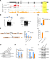
Fig. 5. Bud31 depletion leads to increased retention of the first intron of Cdk2.
A The AS pattern and Bud31 direct binding sites in the Cdk2 pre-mRNA were visualized with IGV using the SMART-seq and the RIP-seq data. The yellow region highlights the AS region and Bud31 binding sites. B Semiquantitative RT-PCR was performed to validate AS events in Cdk2. Percent spliced in (PSI) was quantified (n = 3). C Analysis of the splicing of Cdk2 minigene by semiquantitative RT-PCR. D qPCR was performed to analyze long-to-short isoform ratio of Cdk2 in the testes of P10 Bud31-sKO and littermate control mice. E A splicing reporter assay was performed to assess the splicing of Cdk2 intron 1 from the pre-mRNA molecule as regulated by Bud31 in GC-1 cells (n = 3 biologically independent samples). F RIP-qPCR was performed to validate the interaction between Bud31 and Cdk2 RNA in P14 mouse testes primary cells using the Bud31 antibody and IgG. G RNA pull-down assay showing the interaction between the Cdk2 intron 1 transcript and Bud31 protein. H qPCR was performed to measure Cdk2 expression in testes tissues of P10 Bud31-sKO and littermate control mice. I qPCR analysis of Cdk2 expression in GC-1 cells transfected with control and Bud31 siRNA. Student’s t test was performed, and data are shown as the mean ± SEM of three independent experiments. *P < 0.05, **P < 0.01, ***P < 0.001. J Western blotting analysis of P-Cdk2, Cdk2 and Bud31 levels in testes from P4 Bud31-vKO and control mice. Tubulin was used as the loading control.

References

    1. Song HW, Bettegowda A, Lake BB, Zhao AH, Skarbrevik D, Babajanian E, et al. The homeobox transcription factor RHOX10 drives mouse spermatogonial stem cell establishment. Cell Rep. 2016;17:149–64. doi: 10.1016/j.celrep.2016.08.090. - DOI - PMC - PubMed
    1. Krawetz SA, De Rooij DG, Hedger MP. Molecular aspects of male fertility. International Workshop on Molecular Andrology. EMBO Rep. 2009;10:1087–92. doi: 10.1038/embor.2009.211. - DOI - PMC - PubMed
    1. Feng LX, Chen Y, Dettin L, Pera RA, Herr JC, Goldberg E, et al. Generation and in vitro differentiation of a spermatogonial cell line. Science. 2002;297:392–5. doi: 10.1126/science.1073162. - DOI - PubMed
    1. Oatley JM, Brinster RL. Regulation of spermatogonial stem cell self-renewal in mammals. Annu Rev Cell Dev Biol. 2008;24:263–86. doi: 10.1146/annurev.cellbio.24.110707.175355. - DOI - PMC - PubMed
    1. Song HW, Wilkinson MF. Transcriptional control of spermatogonial maintenance and differentiation. Semin Cell Dev Biol. 2014;30:14–26. doi: 10.1016/j.semcdb.2014.02.005. - DOI - PMC - PubMed

Publication types