. 2022 Sep 16;12(1):15580.
doi: 10.1038/s41598-022-19903-0.

Zinc chelator treatment in crush syndrome model mice attenuates ischemia-reperfusion-induced muscle injury due to suppressing of neutrophil infiltration

Affiliations

Zinc chelator treatment in crush syndrome model mice attenuates ischemia-reperfusion-induced muscle injury due to suppressing of neutrophil infiltration

Yohei Haruta et al. Sci Rep. .

Abstract

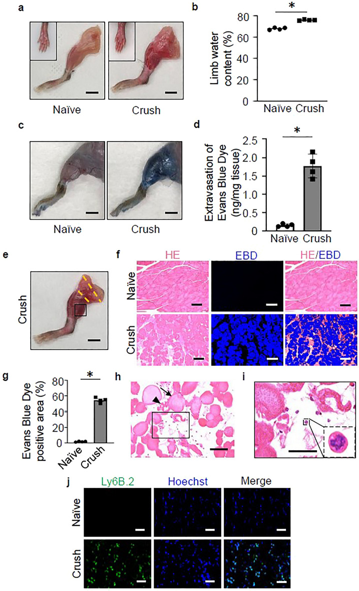
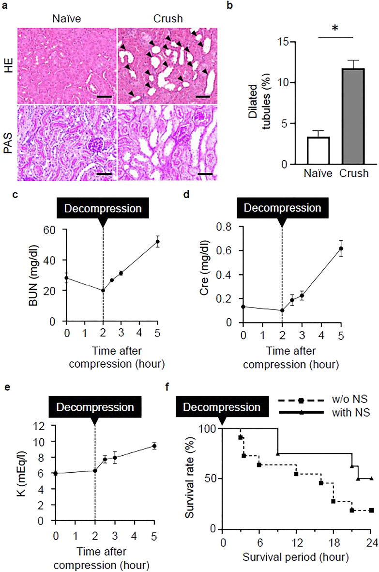
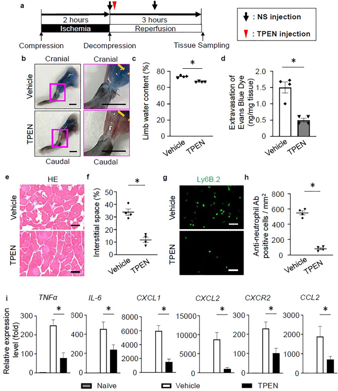
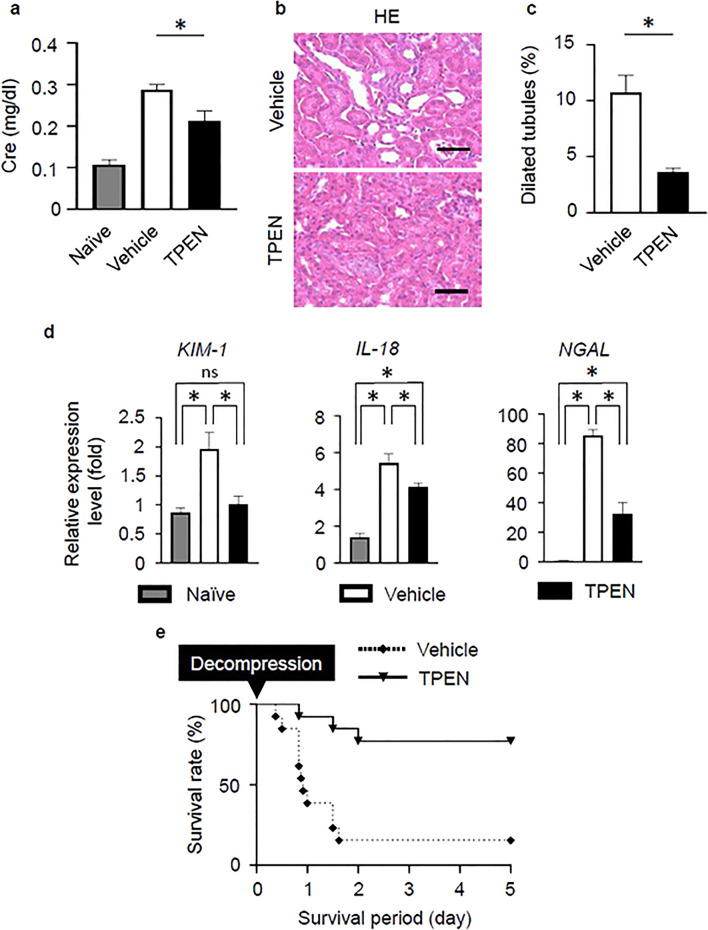
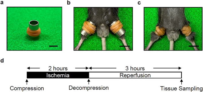
In crush syndrome, massive muscle breakdown resulting from ischemia-reperfusion muscle injury can be a life-threatening condition that requires urgent treatment. Blood reperfusion into the ischemic muscle triggers an immediate inflammatory response, and neutrophils are the first to infiltrate and exacerbate the muscle damage. Since free zinc ion play a critical role in the immune system and the function of neutrophils is impaired by zinc depletion, we hypothesized that the administration of a zinc chelator would be effective for suppressing the inflammatory reaction at the site of ischemia-reperfusion injury and for improving of the pathology of crush syndrome. A crush syndrome model was created by using a rubber tourniquet to compress the bilateral hind limbs of mice at 8 weeks. A zinc chelator N,N,N',N'-tetrakis-(2-pyridylmethyl)-ethylenediamine (TPEN) was administered immediately after reperfusion in order to assess the anti-inflammatory effect of the chelator for neutrophils. Histopathological evaluation showed significantly less muscle breakdown and fewer neutrophil infiltration in TPEN administration group compared with control group. In addition, the expression levels of inflammatory cytokine and chemokine such as IL-6, TNFα, CXCL1, CXCL2, CXCR2, CCL2 in ischemia-reperfusion injured muscle were significantly suppressed with TPEN treatment. Less dilatation of renal tubules in histological evaluation in renal tissue and significantly better survival rate were demonstrated in TPEN treatment for ischemia-reperfusion injury in crush syndrome. The findings of our study suggest that zinc chelators contributed to the resolution of exacerbation of the inflammatory response and attenuation of muscle breakdown in the acute phase after crush syndrome. In addition, our strategy of attenuation of the acute inflammatory reaction by zinc chelators may provide a promising therapeutic strategy not only for crush syndrome, but also for other diseases driven by inflammatory reactions.


Conflict of interest statement

The authors declare no competing interests.

Figures

Figure 1
Figure 1
Vascular hyperpermeability, edematous changes, and neutrophil infiltration was observed in muscles with ischemia–reperfusion injury. (a) Representative images of the hind limbs after removal of the thigh and lower leg subcutaneous tissue with (crush group)/without (naïve group) tourniquet placement. Scale bars: 5 mm. (b) The quantitative analysis of the water content in the hind limbs of both groups at 3 h post-reperfusion (n = 4 per group). P < 0.005, unpaired t test. (c) Representative images of the hind limbs of naïve and crush model mice following the intraperitoneal injection of Evans Blue Dye (EBD). Scale bars: 5 mm. (d) The quantitative analysis of the extravasation of EBD in the hind limbs of both groups at 3 h post-reperfusion (n = 4 per group). P < 0.005, unpaired t test. (e) The muscle tissue was harvested from the gastrocnemius muscle, the region (the square part) distal to the compression site (between the orange broken lines) of the hind limb. Scale bar: 5 mm. (f) Representative images of the EBD uptake in the gastrocnemius muscle of the naïve and crush groups. Overlay image of the fluorescence image (EBD) and bright field image (Hematoxylin Eosin [HE]-stained). Scale bars: 100 μm. (g) The quantitative analysis of the EBD uptake in the gastrocnemius muscle of both groups at 3 h post-reperfusion (n = 4 per group). P < 0.001, unpaired t test. (h) Representative images of HE-stained muscle tissue sections in the crush group. Scale bar: 50 μm. The arrow indicates swelling of myofibers and the arrowhead indicates the breakdown of myofibers. (i) A representative expanded image of the inset of (h). A magnified view of the cell with nucleus between myofibers shown in the additional inset indicated with a broken line. Scale bar: 20 μm. (j) Representative images of muscle tissue specimens analyzed for neutrophils using Ly6B.2 (green) and Hoechst (blue) staining. Scale bars: 20 μm.
Figure 2
Figure 2
Crush syndrome leads to renal injury and fluid therapy improves the survival rate. (a) Representative images of HE and Periodic Acid Schiff (PAS)-stained renal tissue sections at 3 h post-reperfusion in the naïve and crush groups. Scale bars: 50 μm. (b) The quantitative analysis of the dilated tubules in the renal cortex in HE-stained sections from both groups (n = 4 per group). P < 0.05, unpaired t test. (ce) Serum BUN, Cre, and K levels after reperfusion (n = 4 per group). BUN blood urea nitrogen, Cre creatinine, K potassium. (f) The post-reperfusion survival rates with/without normal saline (NS) treatment were calculated using the Log-rank test (Kaplan–Meier method). P < 0.05. CS crush syndrome, NS normal saline.
Figure 3
Figure 3
Zinc chelation therapy in crush syndrome reduces neutrophil infiltration, inflammation, and muscle breakdown. (a) The zinc chelator treatment protocol, N,N,N′,N′-Tetrakis (2-pyridylmethyl) ethylenediamine (TPEN) post-reperfusion. (b) Representative images of the lower legs with vehicle control and TPEN treatment, following the intraperitoneal injection of EBD. The lower legs with the fascia exposed at 3 h post-reperfusion. The orange dotted line indicates the distal end of the compression site of the hind limb. Scale bars: 5 mm. (c) The quantitative analysis of the water content in the hind limbs in the vehicle control and TPEN-treated groups at 3 h post-reperfusion (n = 4 per group). P < 0.05, unpaired t test. (d) The quantitative analysis of the extravasation of EBD in the hind limbs of both groups at 3 h post-reperfusion (n = 4 per group). P < 0.01, unpaired t test. (e) Representative images of HE-stained gastrocnemius muscle sections, the distal side of the compression site from mice in vehicle control and TPEN-treated groups. Scale bars: 50 μm. (f) The quantitative analysis of the interstitial space of the gastrocnemius muscle at mice in vehicle control and TPEN-treated groups (n = 4 per group). P < 0.05, unpaired t test. (g) Representative images of muscle tissue specimens stained with Ly6B.2 (green) in vehicle control and TPEN-treated groups (n = 4 per group). P < 0.005, unpaired t test. Scale bars: 50 μm. (h) The quantitative analysis of the Ly6B.2 (anti-neutrophil antibody)-positive cell count in vehicle control and TPEN-treated groups (n = 4 per group). P < 0.01, unpaired t test. (i) The TNFα, IL-6, CXCL1, CXCL2, CXCR2, CCL2 mRNA expression levels in vehicle control and TPEN-treated groups were compared (n = 4 per group). P < 0.05, unpaired t test.
Figure 4
Figure 4
Zinc chelation therapy improves the renal function and survival rate in crush syndrome. (a) The effects of TPEN treatment on the serum Cre levels at 3 h after reperfusion (n = 4 per group). P < 0.05, unpaired t test. (b) Representative images of HE-stained renal tissue in vehicle control and TPEN-treated groups at 3 h post-reperfusion. Scale bars: 50 μm. (c) The quantitative analysis of the dilated tubules in the renal cortex in HE-stained sections in vehicle control and TPEN-treated groups (n = 4 per group). P < 0.05, unpaired t test. (d) The KIM-1, IL-18, NGAL mRNA expression levels in 3 groups (Naïve, vehicle control, TPEN-treated) were compared (n = 4 per group). P < 0.05, one-way ANOVA with the Tukey–Kramer post hoc test. (e) The post-reperfusion survival rate in vehicle control and TPEN-treated groups (n = 13 per group). P < 0.001, Log-rank test.
Figure 5
Figure 5
Establishment of the mouse model of crush syndrome and the experimental protocol. (a) Equipment for the insertion of the rubber tourniquet. Scale bar: 1 mm. (b) Insertion of the tourniquets by sliding on the pipe. Scale bar: 1 mm. (c) Representative positioning of the rubber tourniquets on both hind limbs. Scale bar: 1 mm. (d) Ischemia of the hind limbs caused by the tourniquet persists for 2 h. The hind limbs are collected at 3 h post-reperfusion (after release of the tourniquet).

References

    1. von Colmers F. Uber die durch das Erbeben in Messina. Arch. fur klin. Chir. 1909;90:701–747.
    1. Bywaters EGL, Beall D, Bywaters GL, Knochel P. Crush injuries with impairment by of renal. J. Am. Soc. Nephrol. 1941;1:427–432.
    1. Murata I, et al. Characterization of systemic and histologic injury after crush syndrome and intervals of reperfusion in a small animal model. J. Trauma Inj. Infect. Crit. Care. 2011;70:1453–1463. doi: 10.1097/TA.0b013e31820ca00a. - DOI - PubMed
    1. Vanholder R, et al. Earthquakes and crush syndrome casualties: Lessons learned from the Kashmir disaster. Kidney Int. 2007;71:17–23. doi: 10.1038/sj.ki.5001956. - DOI - PubMed
    1. Schofield ZV, Woodruff TM, Halai R, Wu MCL, Cooper MA. Neutrophils—A key component of ischemia–reperfusion injury. Shock. 2013;40:463–470. doi: 10.1097/SHK.0000000000000044. - DOI - PubMed

Publication types

MeSH terms