. 2022 Sep 16;13(1):476.
doi: 10.1186/s13287-022-03159-6.

The role of DNA demethylation in liver to pancreas transdifferentiation

Affiliations

The role of DNA demethylation in liver to pancreas transdifferentiation

Adi Har-Zahav et al. Stem Cell Res Ther. .

Abstract

Background: Insulin producing cells generated by liver cell transdifferentiation, could serve as an attractive source for regenerative medicine. The present study assesses the relationship between DNA methylation pTFs induced liver to pancreas transdifferentiation.

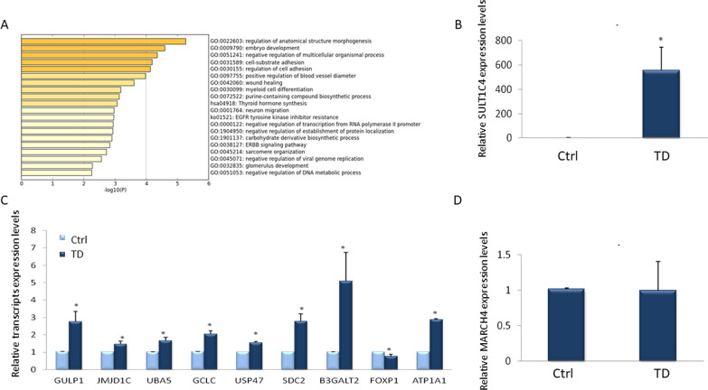
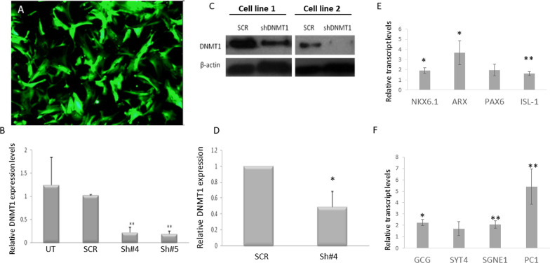
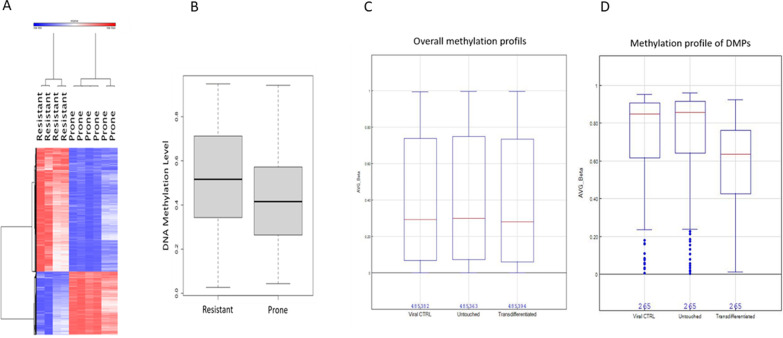
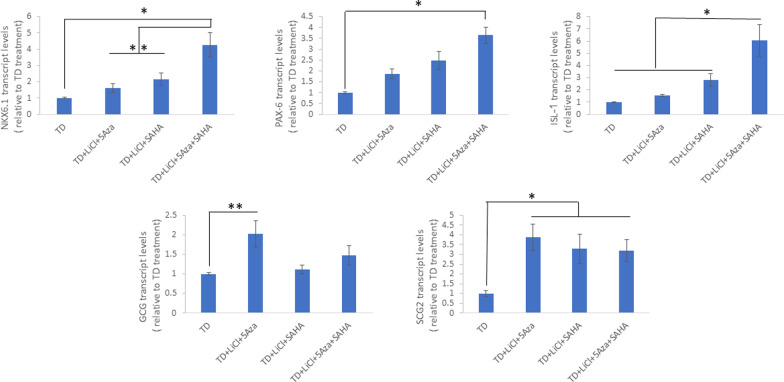
Results: The transdifferentiation process is associated with DNA demethylation, mainly at gene regulatory sites, and with increased expression of these genes. Active inhibition of DNA methylation promotes the pancreatic transcription factor-induced transdifferentiation process, supporting a causal role for DNA demethylation in this process.

Conclusions: Transdifferentiation is associated with global DNA hypomethylation, and with increased expression of specific demethylated genes. A combination of epigenetic modulators may be used to increase chromatin accessibility of the pancreatic transcription factors, thus promoting the efficiency of the developmental process.

Keywords: Cell replacement therapy for diabetes; DNA methylation; Epigenetic modifications; Liver; Pancreas; Pancreatic transcription factors; Transdifferentiation.


Conflict of interest statement

IML is an employee of Orgenesis LTD, SF is employee and consultant of Orgenesis LTD. Interpretation of results, and subsequent submission and publication decisions have been made independent of the sponsors. The authors have no other relevant affiliations or financial involvement with any organization or entity with a financial interest in or financial conflict with the subject matter or materials discussed in the manuscript apart from those disclosed. No writing assistance was utilized in the production of this manuscript.

Figures

Fig. 1
Fig. 1
TD induces DNA de-methylation mainly at enhancer sites. Heat maps of the 192 CpGs differentially hypomethylated (< 10%) at day 6 post TD induction in donor A (A) and in donor B (B), comparing TD cells (TD) to un-treated liver cells (UT) or cells infected with adenoviral control (Ad-β-gal; Ctrl). Distribution of the hypomethylated DMPs in enhancers, in TD cells from donor A (C) and donor B (D). *p Value 4.5e−157 in C; p value = 1.14e−09 in D
Fig. 2
Fig. 2
Activation of gene expression levels of demethylated genes in TD. A Functional analysis of DMPs with methylation differences (< −10%) (adj P value < 0.1). BD Liver cells were induced to transdifferentiate for 6 days. The expression levels of genes with hypermethylated CpG’s (B, C) and genes with no change in DNA methylation (D) were measured by RT-qPCR. Results are presented as average ± SD, n = 6 different donors, p < 0.05
Fig. 3
Fig. 3
TD induces gradual CpG demethylation. A Heat map of top 52 differentially methylated CpGs with delta =  > 0.15 from donor B, at day 2 and day 6 after initiation of TD (adjusted P < 0.05) (Pearson linear correlation). B Liver cells were induced to transdifferentiate for 6 days. Gene expression analysis was performed 2–6 days post trans-differentiation of genes that were demethylated by the treatment: A SDC1 (Syndecan 1), B USP47 (Ubiquitin Specific Peptidase 47), C JmjD1C, (Jumonji Domain Containing 1C) D SULT1C4, E UBA5 (Ubiquitin Like Modifier Activating Enzyme 5), and F GCLC (Glutamate-Cysteine Ligase Catalytic Subunit). Results are the average of RNA extracted from 3 different donors
Fig. 4
Fig. 4
Downregulation of DNMT1 expression promotes pTFs induced TD: DNMT1 expression was knocked-down using specific shRNAs (sh#4 and sh#5) delivered by GFP-expressing lentiviruses, as described in materials and methods: lentivirus infection efficiency was measured by A GFP expression of treated cells at 2 weeks post lentiviral infection. B mRNA levels of DNMT1 at 2 weeks post lentiviral infection. N = 8 p < 0.000003. C Western blot analysis of DNMT1 protein levels in 2 selected cell lines at 2 weeks post infection. D Protein level quantification of DNMT1 at 2 weeks post lentiviral infection, compared to scrambled shRNA delivery. N = 4, p < 0.0006. Stable clones of DNMT1 knocked down liver cells were transdifferentiated by ectopic expression of pTFs. The expression levels of pancreatic specific TFs (E) and pancreatic specific genes (F) was measured 6 days post TD induction by RT-qPCR. Results are presented as average ± SD, n = 4 different donors, *p < 0.01, **p < 0.05
Fig. 5
Fig. 5
DNA demethylation agent 5-AZA promotes pTF-induced TD: liver cells were treated by increasing amounts of 5-AZA (0–4 µM), 48 h before induction of trans-differentiation by ectopic pTF expression. The expression levels of pancreatic specific transcription factors (A) and pancreatic specific genes (B) was measured 6 days post TD induction by RT-qPCR. Results are presented as average ± SD, n = 6 different donors, *p < 0.01, **p < 0.05
Fig. 6
Fig. 6
TD-prone cells display basal DNA hypomethylation compared to TD-resistant liver cells and further undergo demethylation upon pTF-induced TD A Heat map of 6017 CpGs with over 20% differences in resistant (n = 4) and prone cells (n = 6), adjP < 0.05. B Boxplot display of 6017 over 20% DMPs in resistant (n = 4) vs. prone (n = 6) human liver primary cell lines, adjP < 0.05. TD-prone cells underwent the TD protocol, and average global methylation across all probes in Infinium HumanMethylation450 BeadChip Kit was determined (Illumina). C Overall methylation profile of viral control (β-gal) infected cells, untreated cells (untouched), and TD cells. Red lines indicate the average methylation level. D Methylation profile of altered meCgGs. Compared to untreated cells, 265 CpG’s methylation levels were significantly altered in TD cells
Fig. 7
Fig. 7
Significant increase in TD efficiency occurs upon combination of pTFs with epigenetic modifying agents. Liver cells were treated with a combination of soluble factors, as described in the materials and methods section, and infected 48 h later with pTFs. Expression of the pTFs NKX6.1,PAX6, and ISL-1 as well as the pancreatic specific genes GCG, SST and SCG2was analyzed by RT-qPCR. Results are presented as average ± SD, n = 3 different donors, *p < 0.01, **p < 0.05 related to TD alone, ##p < 0.01, #p < 0.05 related to TD + LiCl + 5-AZA + SAHA

References

    1. Graf T. Historical origins of transdifferentiation and reprogramming. Cell Stem Cell. 2011;9(6):504–516. doi: 10.1016/j.stem.2011.11.012. - DOI - PubMed
    1. Ber I, Shternhall K, Perl S, Ohanuna Z, Goldberg I, Barshack I, et al. Functional, persistent, and extended liver to pancreas transdifferentiation. J Biol Chem. 2003;278(34):31950–31957. doi: 10.1074/jbc.M303127200. - DOI - PubMed
    1. Meivar-Levy I, Sapir T, Gefen-Halevi S, Aviv V, Barshack I, Onaca N, et al. Pancreatic and duodenal homeobox gene 1 induces hepatic dedifferentiation by suppressing the expression of CCAAT/enhancer-binding protein beta. Hepatology (Baltimore, MD) 2007;46(3):898–905. doi: 10.1002/hep.21766. - DOI - PubMed
    1. Sapir T, Shternhall K, Meivar-Levy I, Blumenfeld T, Cohen H, Skutelsky E, et al. From the cover: cell-replacement therapy for diabetes: generating functional insulin-producing tissue from adult human liver cells. Proc Natl Acad Sci USA. 2005;102(22):7964–7969. doi: 10.1073/pnas.0405277102. - DOI - PMC - PubMed
    1. Matei IV, Meivar-Levy I, Lixandru D, Dima S, Florea IR, Ilie VM, et al. The effect of liver donors' age, gender and metabolic state on pancreatic lineage activation. Regen Med. 2021;16(1):19–31. doi: 10.2217/rme-2020-0092. - DOI - PubMed

Publication types