. 2022 Sep 16;14(1):131.
doi: 10.1186/s13098-022-00904-y.

Adiponectin reduces apoptosis of diabetic cardiomyocytes by regulating miR-711/TLR4 axis

Affiliations

Adiponectin reduces apoptosis of diabetic cardiomyocytes by regulating miR-711/TLR4 axis

Yu Zuo et al. Diabetol Metab Syndr. .

Abstract

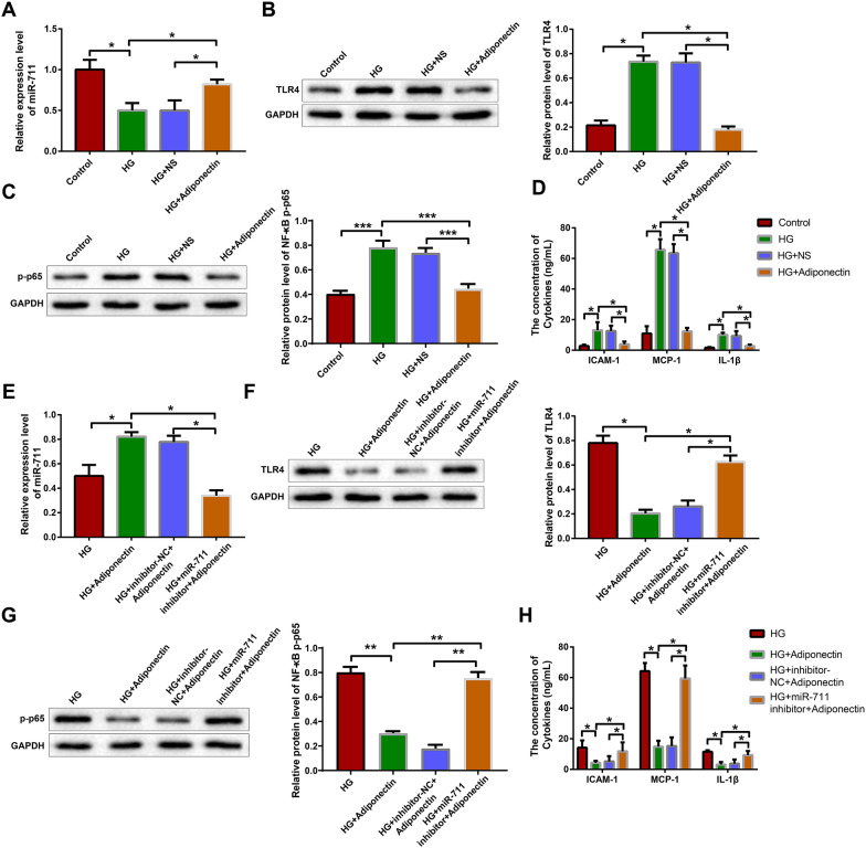
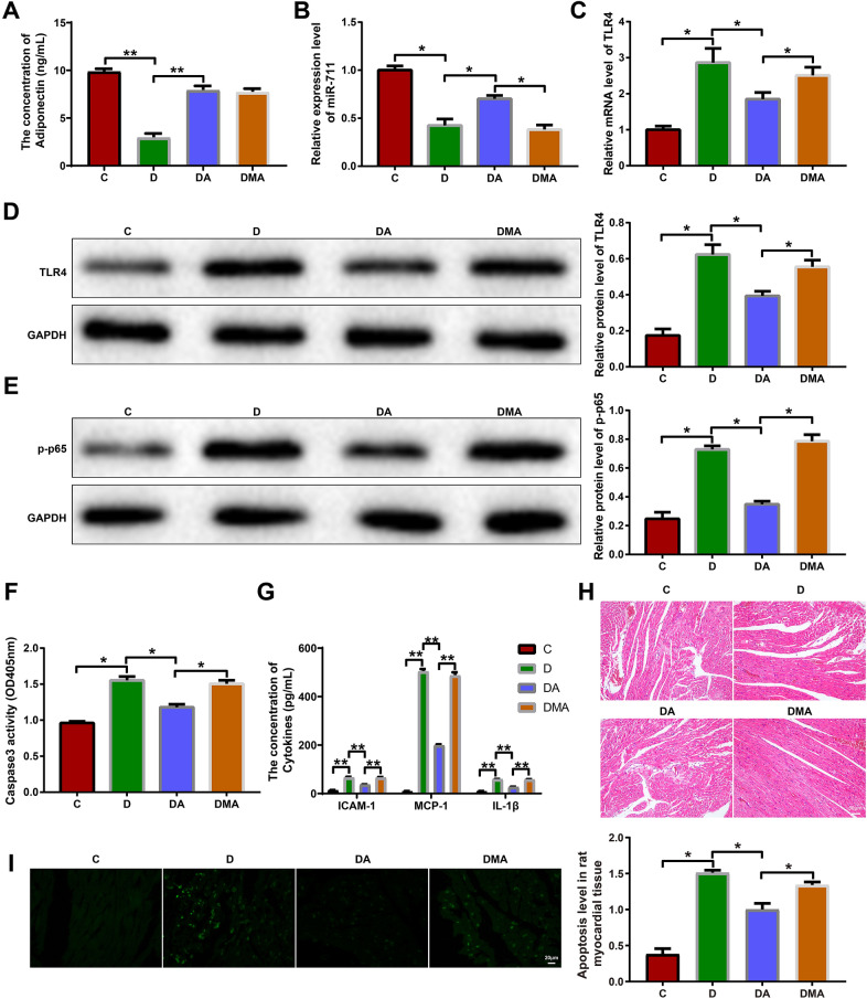
Objective: To investigate the regulation of adiponectin/miR-711 on TLR4/NF-κB-mediated inflammatory response and diabetic cardiomyocyte apoptosis.

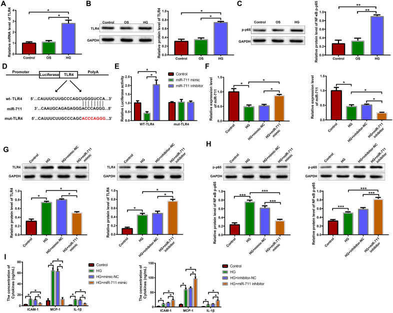
Methods: Diabetes models were established using rats and H9c2 cardiomyocytes. qRT-PCR was used to detect adiponectin, miR-711, and TLR4. MTT, β-galactosidase staining, and flow cytometry were utilized to assess cell viability, senescence, and apoptosis, respectively. The colorimetric method was used to measure caspase-3 activity, DCFH-DA probes to detect ROS, and western blotting to determine the protein levels of Bax, Bcl-2, TLR4, and p-NF-κB p65. ELISA was performed to measure the levels of adiponectin, ICAM-1, MCP-1, and IL-1β. Dual-luciferase reporter system examined the targeting relationship between miR-711 and TLR4. H&E and TUNEL staining revealed myocardial structure and apoptosis, respectively.

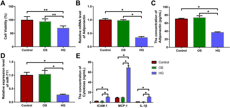
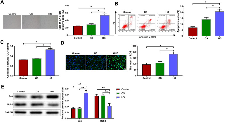
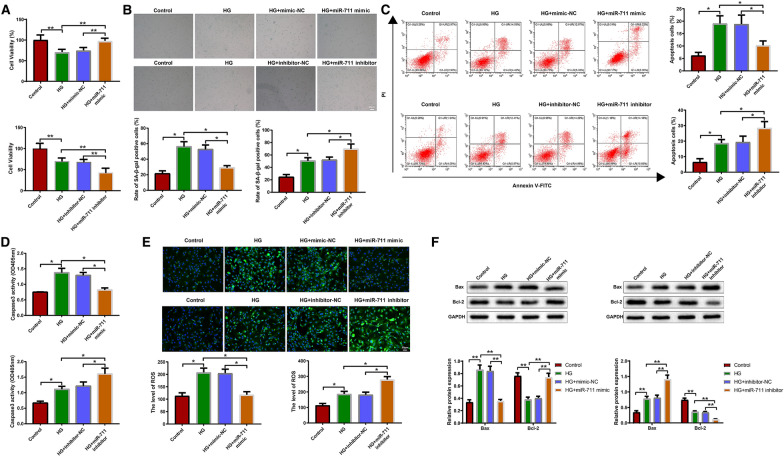
Results: Adiponectin and miR-711 were underexpressed and TLR4/NF-κB signaling pathway was activated in high glucose-treated H9c2 cells. High glucose treatment reduced viability, provoked inflammatory response, and accelerated senescence and apoptosis in H9c2 cells. miR-711 could bind TLR4 mRNA and inactivate TLR4/NF-κB signaling. Adiponectin treatment increased miR-711 expression and blocked TLR4/NF-κB signaling. Adiponectin/miR-711 reduced myocardial inflammation and apoptosis in diabetic rats.

Conclusion: Adiponectin inhibits inflammation and alleviates high glucose-induced cardiomyocyte apoptosis by blocking TLR4/NF-κB signaling pathway through miR-711.

Keywords: Adiponectin; Apoptosis; Cardiomyocyte; Diabetes; TLR4; miRNA-711.


Conflict of interest statement

The authors declare there is no conflict of interest regarding this study.

Figures

Fig. 1
Fig. 1
Adiponectin and miR-711 were underexpressed in high glucose-treated H9c2 cells. H9c2 cells were treated with mannitol or high glucose. A MTT was used to detect cell viability. B qRT-PCR was used to detect the expression of adiponectin in cells. C ELISA was performed to detect the expression of adiponectin in cell culture supernatant. D qRT-PCR was used to detect the expression of miR-711 in cells. E ELISA was performed to detect the expression of ICAM-1, MCP-1, and IL-1β in cell culture supernatant. The data were all measurement data and expressed as mean ± standard deviation. One-way analysis of variance was used for comparisons between multiple groups and Tukey's test for post-hoc multiple comparisons. * indicates P < 0.05. The experiments were repeated thrice. OS osmotic, HG high glucose, MTT, 3-(4,5-dimethylthiazol-2-yl)-2,5-diphenyltetrazolium bromide, qRT-PCR quantitative real-time polymerase chain reaction, ELISA enzyme-linked immunosorbent assay, ICAM intercellular adhesion molecule, MCP monocyte chemotactic protein, IL interleukin
Fig. 2
Fig. 2
High glucose treatment impacts on the biological behaviors of H9c2 cells. H9c2 cells were treated with mannitol or high glucose. A β-galactosidase staining was used to reveal cell senescence. B Flow cytometry was used to analyze cell apoptosis. C The colorimetric method was used to detect the activity of caspase-3 in cells. D DCFH-DA probes were used to detect ROS in cells. E Western blotting was used to detect Bax and Bcl-2 in cells. The data were all measurement data and expressed as mean ± standard deviation. One-way analysis of variance was used for comparisons between multiple groups and Tukey's test for post-hoc multiple comparisons. * indicates P < 0.05. The experiments were repeated thrice. OS osmotic, HG high glucose, DCFH-DA, 2′,7′-dichlorodihydrofluorescein diacetate, ROS reactive oxygen species, Bax Bcl-2-associated X protein, Bcl-2 B-cell lymphoma-2
Fig. 3
Fig. 3
miR-711 negatively regulates TLR4 and reduces high glucose-provoked inflammatory responses of H9c2 cells. A qRT-PCR was used to detect the expression of TLR4 mRNA in H9c2 cells treated with mannitol or high glucose. Western blotting was used to detect the expression of TLR4 protein B and p-NF-κB p65 protein C in H9c2 cells treated with mannitol or high glucose. D TargetScan database predicted the binding sites between miR-711 and TLR4 mRNA and mutations were designed. E Dual-luciferase reporter assay was performed to verify the binding between miR-711 and TLR4 mRNA. Next, high glucose-treated H9c2 cells were transfected with mimic-NC, miR-711 mimic, inhibitor-NC or miR-711 inhibitor. F qRT-PCR was used to detect the expression of miR-711 in the transfected cells. Western blotting was used to detect the expression of TLR4 protein G and p-NF-κB p65 protein H in the transfected cells. I ELISA was performed to detect the concentrations of ICAM-1, MCP-1, and IL-1β in cell culture supernatant. The data were all measurement data and expressed as mean ± standard deviation. One-way analysis of variance was used for comparisons between multiple groups and Tukey’s test for post-hoc multiple comparisons. * indicates P < 0.05. The experiments were repeated thrice. OS osmotic, HG high glucose, NC negative control, TLR4 toll-like receptor 4, qRT-PCR quantitative real-time polymerase chain reaction, ELISA enzyme-linked immunosorbent assay, ICAM intercellular adhesion molecule, MCP monocyte chemotactic protein, IL interleukin
Fig. 4
Fig. 4
miR-711 affects the biological behaviors of high glucose-treated H9c2 cells by regulating TLR4/NF-κB. High glucose-treated H9c2 cells were transfected with mimic-NC, miR-711 mimic, inhibitor-NC or miR-711 inhibitor. A MTT assay was performed to assess cell viability. B β-galactosidase staining was used to reveal cell senescence. C Flow cytometry was used to analyze cell apoptosis. D The colorimetric method was used to detect the activity of caspase-3 in cells. E DCFH-DA probes were used to detect ROS in cells. F Western blotting was used to detect Bax and Bcl-2 in cells. The data were all measurement data and expressed as mean ± standard deviation. One-way analysis of variance was used for comparisons between multiple groups and Tukey's test for post-hoc multiple comparisons. * indicates P < 0.05. The experiments were repeated thrice. HG high glucose, NC negative control, TLR4 toll-like receptor 4, NF-κB nuclear factor κB, MTT, 3-(4,5-dimethylthiazol-2-yl)-2,5-diphenyltetrazolium bromide; DCFH-DA, 2′,7′-dichlorodihydrofluorescein diacetate; ROS reactive oxygen species, Bax Bcl-2-associated X protein; Bcl-2 B-cell lymphoma-2
Fig. 5
Fig. 5
Adiponectin regulates TLR4 inflammatory signaling through miR-711. H9c2 cells cultured in high glucose medium were treated with NS or adiponectin: A qRT-PCR was used to detect the expression of miR-711. Western blotting was used to detect the expression of TLR4 protein B and p-NF-κB p65 protein (C). D ELISA was performed to detect the levels of ICAM-1, MCP-1, and IL-1β in cell culture supernatant. Diabetic H9c2 cells were treated adiponectin and transfected with miR-711 inhibitor or inhibitor-NC: E qRT-PCR was used to detect the expression of miR-711. Western blotting was used to detect the expression of TLR4 protein F and p-NF-κB p65 protein (G). H ELISA was performed to detect the levels of ICAM-1, MCP-1, and IL-1β in cell culture supernatant. The data were all measurement data and expressed as mean ± standard deviation. One-way analysis of variance was used for comparisons between multiple groups and Tukey’s test for post-hoc multiple comparisons. * indicates P < 0.05. The experiments were repeated thrice. HG high glucose, NS normal saline, NC negative control, TLR4 toll-like receptor 4, qRT-PCR quantitative real-time polymerase chain reaction, ELISA enzyme-linked immunosorbent assay, ICAM intercellular adhesion molecule, MCP monocyte chemotactic protein, IL interleukin
Fig. 6
Fig. 6
Adiponectin/miR-711 regulates myocardial inflammation and apoptosis in diabetic rats. Diabetic rats were injected with adiponectin or miR-711 + adiponectin. A ELISA was used to determine the contents of serum adiponectin. Quantitative real-time polymerase chain reaction was used to detect the expression of miR-711 B and TLR4 mRNA C in the blood of the rats. Western blotting was used to detect TLR4 D and p-NF-κB p65 E in the myocardium of the rats. F The colorimetric method was used to detect the activity of caspase-3 in the myocardium of the rats. G ELISA was performed to detect the concentrations of serum ICAM-1, MCP-1, and IL-1β. H Hematoxylin and eosin staining was used to reveal myocardial structure. I TUNEL assay was used to reveal myocardial apoptosis. n = 3. The data were all measurement data and expressed as mean ± standard deviation. One-way analysis of variance was used for comparisons between multiple groups and Tukey’s test for post-hoc multiple comparisons. * indicates P < 0.05. C control group, D diabetes group, DA diabetes + adiponectin group, DMA diabetes + miR-711 inhibitor + adiponectin group, TLR4 toll-like receptor 4, ICAM intercellular adhesion molecule, MCP monocyte chemotactic protein, IL interleukin, TUNEL terminal deoxynucleotidyl transferase-mediated dUTP-biotin nick end labeling

References

    1. Petersmann A, Muller-Wieland D, Muller UA, Landgraf R, Nauck M, Freckmann G, Heinemann L, Schleicher E. Definition, classification and diagnosis of diabetes mellitus. Exp Clin Endocrinol Diabetes. 2019;127:S1–S7. doi: 10.1055/a-1018-9078. - DOI - PubMed
    1. Schmidt AM. Highlighting diabetes mellitus: the epidemic continues. Arterioscler Thromb Vasc Biol. 2018;38:e1–e8. doi: 10.1161/ATVBAHA.117.310221. - DOI - PMC - PubMed
    1. Zheng Y, Ley SH, Hu FB. Global aetiology and epidemiology of type 2 diabetes mellitus and its complications. Nat Rev Endocrinol. 2018;14:88–98. doi: 10.1038/nrendo.2017.151. - DOI - PubMed
    1. Cole JB, Florez JC. Genetics of diabetes mellitus and diabetes complications. Nat Rev Nephrol. 2020;16:377–390. doi: 10.1038/s41581-020-0278-5. - DOI - PMC - PubMed
    1. Kenny HC, Abel ED. Heart failure in type 2 diabetes mellitus. Circ Res. 2019;124:121–141. doi: 10.1161/CIRCRESAHA.118.311371. - DOI - PMC - PubMed