. 2022 Sep;27(9):097002.
doi: 10.1117/1.JBO.27.9.097002.

Near-infrared diffuse in vivo flow cytometry

Affiliations

Near-infrared diffuse in vivo flow cytometry

Joshua Pace et al. J Biomed Opt. 2022 Sep.

Abstract

Significance: Diffuse in vivo flow cytometry (DiFC) is an emerging technique for enumerating rare fluorescently labeled circulating cells noninvasively in the bloodstream. Thus far, we have reported red and blue-green versions of DiFC. Use of near-infrared (NIR) fluorescent light would in principle allow use of DiFC in deeper tissues and would be compatible with emerging NIR fluorescence molecular contrast agents.

Aim: We describe the design of a NIR-DiFC instrument and demonstrate its use in optical flow phantoms in vitro and in mice in vivo.

Approach: We developed an improved optical fiber probe design for efficient collection of fluorescence from individual circulating cells and efficient rejection of instrument autofluorescence. We built a NIR-DiFC instrument. We tested this with NIR fluorescent microspheres and cell lines labeled with OTL38 fluorescence contrast agent in a flow phantom model. We also tested NIR-DiFC in nude mice injected intravenously with OTL38-labeled L1210A cells.

Results: NIR-DiFC allowed detection of circulating tumor cells (CTCs) in flow phantoms with mean signal-to-noise ratios (SNRs) of 19 to 32 dB. In mice, fluorescently labeled CTCs were detectable with mean SNR of 26 dB. NIR-DiFC also exhibited orders significantly lower autofluorescence and false-alarm rates than blue-green DiFC.

Conclusions: NIR-DiFC allows use of emerging NIR contrast agents. Our work could pave the way for future use of NIR-DiFC in humans.

Keywords: contrast agents; diffuse fluorescence; diffuse in vivo flow cytometry; near-infrared light.


Figures

Fig. 1
Fig. 1
The NIR-DiFC instrument: (a) optical schematic and (b) photograph with main components labeled. (c) Schematic of the newly designed NIR-DiFC fiber probe bundle; see main text for component details. (d), (e) Representative NIR-DiFC data measured from probes 1 and 2, respectively, before postprocessing. As discussed in detail in the text, this requires background subtraction (BS) and peak matching (PM). (f) Postprocessed data after BS and identification of forward-matched peak (green arrowheads) between the two probes.
Fig. 2
Fig. 2
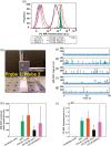
(a) The brightness of OTL38-labeled FR+ cells and JGLI microspheres with flow cytometry. (b) NIR-DIFC with a tissue mimicking flow phantom model. Representative NIR-DiFC data are shown after signal processing for (c) control suspensions of unlabeled L1210A cells, showing no false positive counts, (d) JGLI microspheres, OTL38-labeled (e) L1210A cells, (f) SKOV-3 cells, and (g) IGROV-1 cells. Each peak (arrowhead symbols) represents a forward-matched detection. (h) Comparison of the mean amplitude (intensity) of detected peaks for each sample measured with NIR-DiFC, compared to that measured using (i) flow cytometry, showing good general agreement with NIR-DiFC fluorescence measurements.
Fig. 3
Fig. 3
In vivo testing of NIR-DiFC. (a) Fiber probes were placed on the surface of the mouse tail approximately above the ventral caudal artery. (b) Representative NIR-DiFC data measured from a mouse injected intravenously with OTL38-labeled L1210A cells. Each green arrowhead is an arterial matched cell (traveling in artery), each orange arrowhead is a venous matched cell (traveling in vein), and each yellow-labeled peak represents a detected unmatched cell (traveling in capillary). (c) Representative NIR-DiFC data from a control mouse injected with L1210A. (d) Representative NIR-DiFC data from an uninjected control mouse. (e), (f) Cell detected sequentially in probes 1 and 2, indicating a cell traveling in the arterial direction. (g), (h) Cell detected sequentially in probes 2 and 1, indicating a cell traveling in the venous direction. (i) The mean cell detection rate over a 1-h scan indicating that cell numbers were approximately stable in circulation. Range bars show the minimum and maximum values (N=3). (j) We counted L1210A cells still in circulation following NIR-DiFC. Representative flow cytometry data from blood samples. As described in the text, L1210A cells were prelabeled with both CFSE (green) and OTL38 (NIR) prior to injection appearing as the double-labeled population (Q2).
Fig. 4
Fig. 4
We studied the effect of injection of 2.5  μg OTL38 on the background NIR-DiFC signal and noise following injection. (a) The background increased by a factor of 5.5, 3 h post OTL38 injection compared to the preinjection. By 24 h, the background was near baseline. (b) Likewise the noise (standard deviation of the background), followed a similar trend and returned close to baseline by 24 h. Considering the (c) SNR of detected OTL38-labeled L1210A cells as in Fig. 3, we estimated the effect of the increased noise on the SNR (d) 3 h, (e) 6 h, (f) 9 h, (g) 12 h, and (h) 24 h post-OTL38 injection. The red vertical line at 13.9 dB represents the cell detection threshold, and the percentage of detectable cells compared to baseline as well as the mean SNR is indicated on each plot.
Fig. 5
Fig. 5
(a) Example breathing artifacts measured with our blue-green b-DiFC instrument. (b) These generally can be corrected by simply adjusting the b-DiFC probe position on the skin, although this causes loss of time. (c) In contrast, these motion (breathing) artifacts were not observed with NIR-DiFC measurements. (d) Use of NIR laser light for DiFC resulted in significantly lower tissue autofluorescence than blue light. (e) In practice, this resulted in a reduction in DiFC FAR.

References

    1. Mehlen P., Puisieux A., “Metastasis: a question of life or death,” Nat. Rev. Cancer 6(6), 449–458 (2006). 10.1038/nrc1886 - DOI - PubMed
    1. Steeg P. S., Theodorescu D., “Metastasis: a therapeutic target for cancer,” Nat. Clin. Pract. Oncol. 5(4), 206–219 (2008). 10.1038/ncponc1066 - DOI - PMC - PubMed
    1. Gupta G. P., Massagué J., “Cancer metastasis: building a framework,” Cell 127(4), 679–695 (2006). 10.1016/j.cell.2006.11.001 - DOI - PubMed
    1. Pantel K., Speicher M. R., “The biology of circulating tumor cells,” Oncogene 35(10), 1216–1224 (2016). 10.1038/onc.2015.192 - DOI - PubMed
    1. Alix-Panabieres C., Pantel K., “Circulating tumor cells: liquid biopsy of cancer,” Clin. Chem. 59(1), 110–118 (2013). 10.1373/clinchem.2012.194258 - DOI - PubMed

Publication types