Review
. 2022 Oct;21(10):922-936.
doi: 10.1016/S1474-4422(22)00188-0.

Towards prevention of diabetic peripheral neuropathy: clinical presentation, pathogenesis, and new treatments

Affiliations
Review

Towards prevention of diabetic peripheral neuropathy: clinical presentation, pathogenesis, and new treatments

Melissa A Elafros et al. Lancet Neurol. 2022 Oct.

Abstract

Diabetic peripheral neuropathy (DPN) occurs in up to half of individuals with type 1 or type 2 diabetes. DPN results from the distal-to-proximal loss of peripheral nerve function, leading to physical disability and sometimes pain, with the consequent lowering of quality of life. Early diagnosis improves clinical outcomes, but many patients still develop neuropathy. Hyperglycaemia is a risk factor and glycaemic control prevents DPN development in type 1 diabetes. However, glycaemic control has modest or no benefit in individuals with type 2 diabetes, probably because they usually have comorbidities. Among them, the metabolic syndrome is a major risk factor for DPN. The pathophysiology of DPN is complex, but mechanisms converge on a unifying theme of bioenergetic failure in the peripheral nerves due to their unique anatomy. Current clinical management focuses on controlling diabetes, the metabolic syndrome, and pain, but remains suboptimal for most patients. Thus, research is ongoing to improve early diagnosis and prognosis, to identify molecular mechanisms that could lead to therapeutic targets, and to investigate lifestyle interventions to improve clinical outcomes.


Conflict of interest statement

Conflict of interest

MAE has nothing to disclose. HA has nothing to disclose. MGS has nothing to disclose. VV has nothing to disclose. DLB reports grants from AstraZeneca, grants from Lilly, grants from Diabetes UK, during the conduct of the study; DLB has acted as a consultant on behalf of Oxford Innovation for Amgen, Bristows, LatigoBio, GSK, Ionis, Lilly, Olipass, Orion, Regeneron and Theranexus over the last 2 years, outside the submitted work; in addition, DLB has a patent application ‘a method for the treatment or prevention of pain, or excessive neuronal activity, or epilepsy’ Application No. 16/337,428 pending. BCC reports personal fees from Medical legal work, grants from Veterans Affairs, personal fees from DynaMed, grants and personal fees from American Academy of Neurology, personal fees from Vaccine Injury Compensation Program, outside the submitted work. ELF has nothing to disclose.

Figures
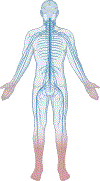
Figure 1.
Figure 1.. DPN presentation
The most common manifestation of DPN is a distal symmetric polyneuropathy, which manifests in the lower limbs first followed by the upper limbs in a ‘stocking-glove’ configuration. DPN symptoms and signs start in the toes (darker pink), which progress proximally towards the calves, at which point, nerve injury initiates in the fingers and moves up to encompass the hands (lighter pink).
Figure 2.
Figure 2.. Pain medication selection protocol for patients with painful DPN. Adapted from Callaghan et al. JAMA, 2020.
Tiered approach to pain management in patients with painful DPN considers. All medications, tricyclic antidepressants (TCAs), serotonin norepinephrine reuptake inhibitors (SNRIs), gabapentinoids (e.g., gabapentin, pregabalin), and sodium channel blockers (e.g., lidocaine) have similar effect sizes. Treatment selection, therefore, should focus on alternative parameters, e.g., tolerability, cost etc. Created with BioRender.com.
Figure 3.
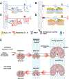
Figure 3.. Neuron anatomy and DPN pathophysiology
(A) Peripheral neuron cell bodies are only microns in width, but axons are up to several feet in length. For normal nerve function, neurons traffic mitochondria from the cell body along the axons to areas of high energy demand. Failure to traffic mitochondria results in energy failure at axon termini followed by distal (pink) -to-proximal (blue) nerve injury, which underlies the stocking-glove pattern of clinical signs and symptoms. (B) Homeostatic conditions (blue), healthy nerve: Axons take up glucose (GLU) in an insulin-independent manner through glucose transporter 3 (GLUT3; green transporter), which is metabolized to pyruvate (PYR) in the cytoplasm and further to ATP in mitochondria. Schwann cells take up glucose in an insulin-dependent manner through GLUT1 (blue transporter); part of the glucose is metabolized for energy use by Schwann cells, part is metabolized to pyruvate then lactate (LAC) for transport to axons through monocarboxylate transporter 1 (MCT1; grey, on Schwann cells) then MCT2 (purple, on axons). Schwann cells take up fatty acids (FA) using fatty acid transporter/ translocase/ binding protein (brown), primarily for myelin production. Mitochondria (blue) are trafficked in the anterograde and retrograde directions to areas of high energy demand. Mitochondria generate a mitochondrial membrane potential for ATP production, powering Na+/K+ ATPase channel activity (pink; purple spheres are cations). Mitochondrial biogenesis occurs to replenish dysfunctional mitochondria (brown), which are eliminated by mitophagy. Pathologic conditions (pink), DPN nerve: Hyperglycemia increases glucose flux (wider arrow) into axons through GLUT3; excess glucose that is not metabolized by glycolysis enters or activates other pathways, e.g., polyol, advanced glycation end products (AGEs), and protein kinase C (PKC) pathways. Downstream, this triggers inflammation and disrupts Na+/K+ ATPase channel activity (no cations pumped, stop sign). Insulin resistance (IR) impairs insulin-dependent GLUT1-facilitated glucose uptake by Schwann cells, disrupting MCT-mediated fuel transport to axons (stop sign) and possibly the balance with lipid metabolism and myelin production. Hyperglycemia and hyperlipidemia also enhance Schwann cell oxidative stress and apoptosis. Hyperlipidemia disrupts axonal mitochondrial biogenesis and trafficking, lowering the fraction and velocity of motile mitochondria. Palmitate also depolarizes axonal mitochondria and reduce their ability to dissipate mitochondrial membrane potential to meet physiological energy demand. Mitophagy is also impaired. Created with BioRender.com.
Figure 4.
Figure 4.. DPN pain mechanisms
(A) Healthy unmyelinated C fiber: Healthy unmyelinated C fiber (blue) with free nerve endings embedded in skin. Inset: Voltage-gated sodium channels (e.g., Nav1.7, 1.8; yellow channel) and ligand-gated channels (e.g., TRPA1; purple channel) transmit an afferent signal if the stimulus (asterisk) is above the channel threshold. (B) Neuropathic unmyelinated C fiber: Unmyelinated C fiber with distal-to-proximal length-dependent neurodegeneration (red) starting in the free nerve endings. Inset: Channel modification by reactive metabolites, e.g., methylglyoxal, reactive species (green oval), or channel mutations (red shaded circle) can lower the threshold potential. Less intense stimulus (asterisk) now falls below the channel threshold, causing neuronal hyperexcitability. (C) Healthy myelinated fiber: Healthy myelinated fiber (blue) with encapsulated nerve ending embedded in skin. Inset: Voltage-gated potassium (Kv; green channel, located to the juxtaparanode) and sodium (Nav; yellow, located to the nodes) channels propagate signals along nodes of Ranvier by saltatory conduction. (D) Neuropathic myelinated fiber: Myelinated fiber with distal-to-proximal length-dependent neurodegeneration (red) starting in the encapsulated nerve ending. Inset: Kv channels expression level drops, leading to hyperexcitability. (E) Simplified schematic of CNS pain mechanisms. Top panel: Ascending pathways of CNS dysfunction arising from peripheral to central pain signal amplification (red arrows). Sensory dorsal root ganglion (DRG; yellow) neurons carrying afferent signals connect with CNS neurons within the spinal cord dorsal horn, which relay within the brainstem to the brain. Different neuron colors (red, green, blue) represent various ascending pain pathways. Bottom panel: Descending pathways of CNS dysfunction arising from brain to spinal cord pain signal amplification (red arrows). Different neuron colors (pink, purple, brown) represent various descending pain pathways. CNS pain pathways give rise to autonomic dysfunction and anxiety, depression, and disturbed sleep. Created with BioRender.com.

Comment in

Similar articles

Cited by

References

    1. Saeedi P, Petersohn I, Salpea P, et al. Global and regional diabetes prevalence estimates for 2019 and projections for 2030 and 2045: Results from the International Diabetes Federation Diabetes Atlas, 9(th) edition. Diabetes Res Clin Pract 2019; 157: 107843. - PubMed
    1. Feldman EL, Callaghan BC, Pop-Busui R, et al. Diabetic neuropathy. Nat Rev Dis Primers 2019; 5(1): 41. - PubMed
    1. Hicks CW, Wang D, Matsushita K, Windham BG, Selvin E. Peripheral Neuropathy and All-Cause and Cardiovascular Mortality in U.S. Adults : A Prospective Cohort Study. Ann Intern Med 2021; 174(2): 167–74. - PMC - PubMed
    1. Callaghan BC, Little AA, Feldman EL, Hughes RA. Enhanced glucose control for preventing and treating diabetic neuropathy. Cochrane Database Syst Rev 2012; 6: CD007543. - PMC - PubMed
    1. Pop-Busui R, Boulton AJ, Feldman EL, et al. Diabetic Neuropathy: A Position Statement by the American Diabetes Association. Diabetes Care 2017; 40(1): 136–54. - PMC - PubMed

Publication types

MeSH terms