. 2022 Sep 17;26(1):45.
doi: 10.1186/s40824-022-00293-3.

The triply periodic minimal surface-based 3D printed engineering scaffold for meniscus function reconstruction

Affiliations

The triply periodic minimal surface-based 3D printed engineering scaffold for meniscus function reconstruction

Lan Li et al. Biomater Res. .

Abstract

Background: The meniscus injury is a common disease in the area of sports medicine. The main treatment for this disease is the pain relief, rather than the meniscal function recovery. It may lead to a poor prognosis and accelerate the progression of osteoarthritis. In this study, we designed a meniscal scaffold to achieve the purposes of meniscal function recovery and cartilage protection.

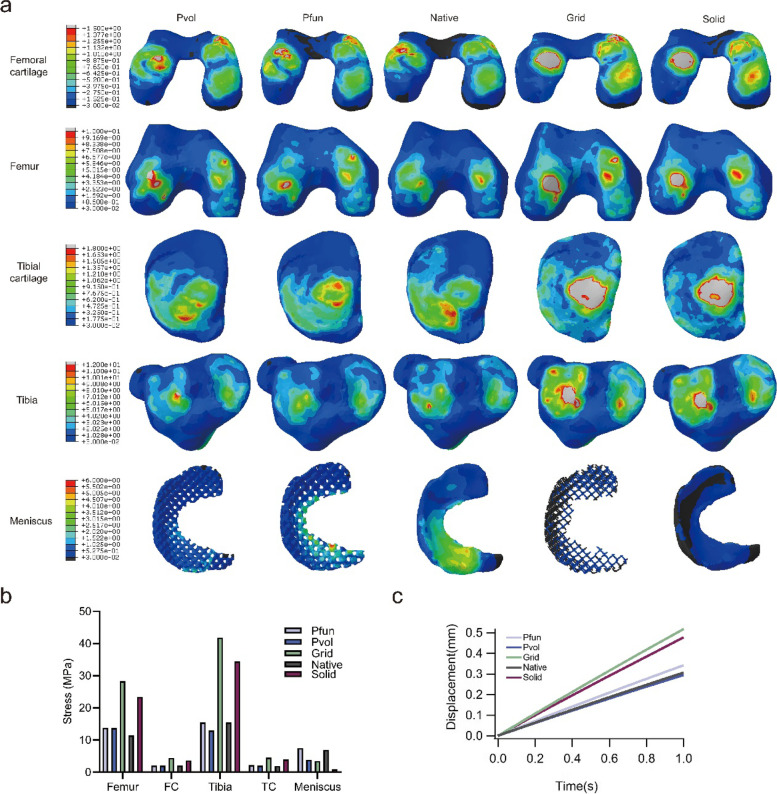
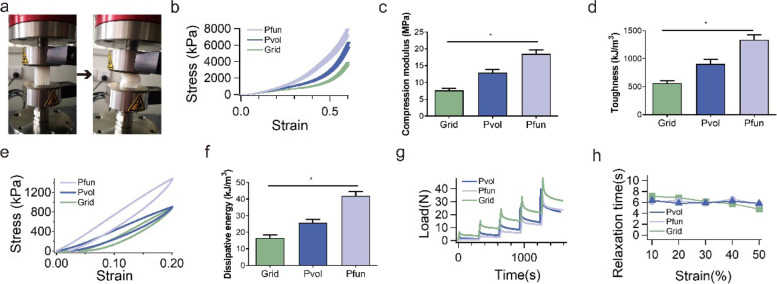
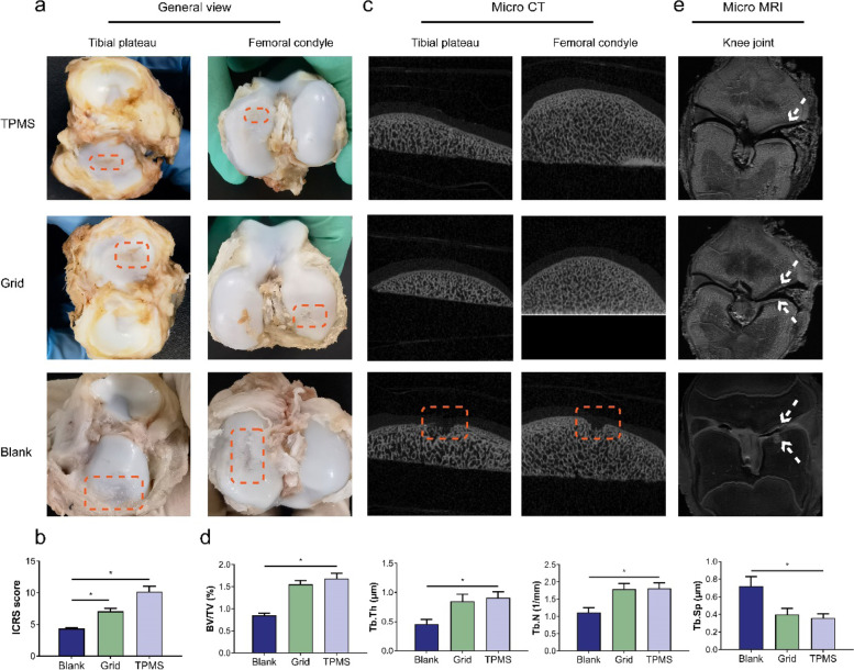
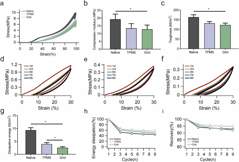
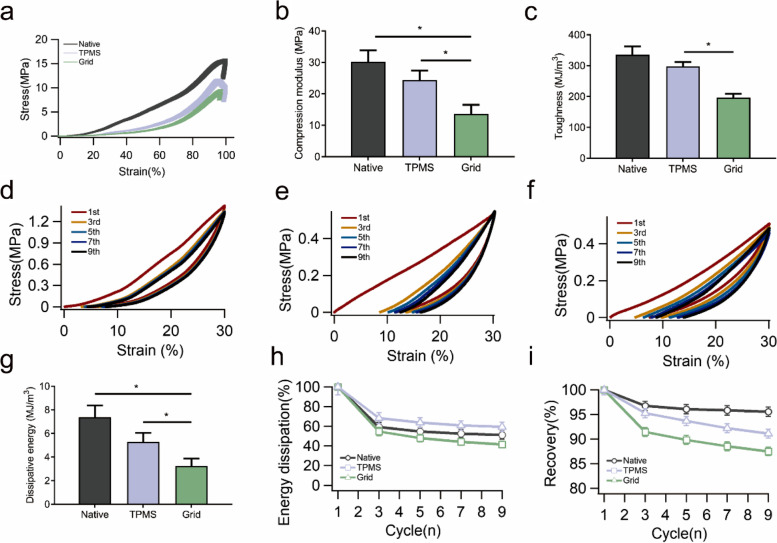
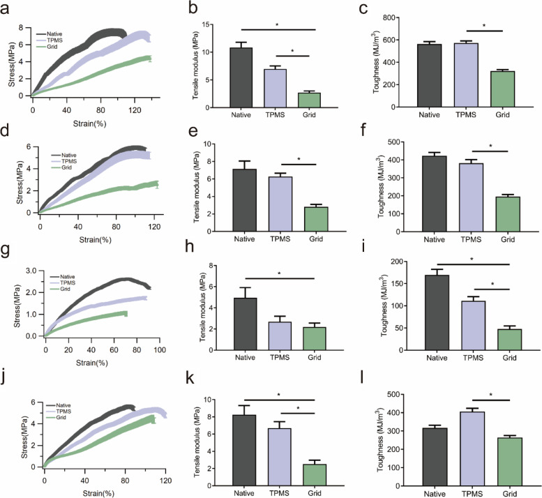
Methods: The meniscal scaffold was designed using the triply periodic minimal surface (TPMS) method. The scaffold was simulated as a three-dimensional (3D) intact knee model using a finite element analysis software to obtain the results of different mechanical tests. The mechanical properties were gained through the universal machine. Finally, an in vivo model was established to evaluate the effects of the TPMS-based meniscal scaffold on the cartilage protection. The radiography and histological examinations were performed to assess the cartilage and bony structures. Different regions of the regenerated meniscus were tested using the universal machine to assess the biomechanical functions.

Results: The TPMS-based meniscal scaffold with a larger volume fraction and a longer functional periodicity demonstrated a better mechanical performance, and the load transmission and stress distribution were closer to the native biomechanical environment. The radiographic images and histological results of the TPMS group exhibited a better performance in terms of cartilage protection than the grid group. The regenerated meniscus in the TPMS group also had similar mechanical properties to the native meniscus.

Conclusion: The TPMS method can affect the mechanical properties by adjusting the volume fraction and functional periodicity. The TPMS-based meniscal scaffold showed appropriate features for meniscal regeneration and cartilage protection.

Keywords: Cartilage protection; Mechanical properties; Meniscal scaffold; Triply periodic minimal surface.


Conflict of interest statement

None.

Figures

Fig. 1
Fig. 1
The diagram of FEA model and porous meniscal scaffold. a The 3D knee model used in the FEA. b Three porous meniscal scaffolds were used in the FEA
Fig. 2
Fig. 2
The results of the FEA. a The stress nephogram on bone, cartilage, and meniscus in the five groups. The color from deep blue to red represented the stress changing from small to large, the gray represented the stress exceeding the threshold in the legend. b The peak stress applied to the main objects of the knee model. c The displacement of meniscal extrusion
Fig. 3
Fig. 3
The results of the in vitro compression tests. a The process of the compression test. b The stress–strain curve of the compression test. c The compression modulus in the three scaffolds. d The toughness in the three scaffolds. e The stress–strain curve of the single-cycle compression at 20% strain. f The dissipative energy for the hysteresis curve. g The curve of the stress-relaxation test. h The relaxation time scale in the three scaffolds. All the tests were repeated for 3 times
Fig. 4
Fig. 4
The general view and radiographic results of the knee joint after 3 months of implantation, n = 5. a General view of the tibial plateau and femoral condyle, the square indicated the lesion cartilage of each group. b The ICRS scores in the three groups, *P < 0.05. c The micro-CT scan images in the three groups, the square indicated the lesion region of subchondral bone in the blank group. d The trabecular parameters in the three groups, *P < 0.05. e The micro-MR images in the three groups, the arrows indicated the cartilage injury in the TPMS and grid group, and the cartilage and subchondral bone defect in the blank group
Fig. 5
Fig. 5
Histological results of the cartilage and meniscus, n = 5. a The histological staining of femoral cartilage in the three groups, the arrows indicated the poorly recovered region at cartilage layer, the squares indicated the obviously defect region at cartilage and subchondral bone, scale bar: 500 μm. b The histological staining of tibial cartilage in the three groups, the arrows indicated the poorly recovered region at cartilage layer, the squares indicated the obviously defect region at cartilage and subchondral bone, scale bar: 500 μm. c The histological staining of meniscus in the three groups, the regenerated region were indicated by the square, scale bar: 1000 μm
Fig. 6
Fig. 6
The results of the compression tests in the inner region, n = 3. a The stress–strain curve of the compression test. b The compression modulus in the three scaffolds, *P < 0.05. c The toughness in the three scaffolds, *P < 0.05. d Five compression-relaxation cycles in the native meniscus. e Five compression-relaxation cycles in the TPMS group. f Five compression-relaxation cycles in the grid group. g The dissipative energy of the 1st cycle in three groups, *P < 0.05. h The percentage of dissipated energy in nine continuous compression-relaxation cycles. i The recovery percentage in nine continuous compression-relaxation cycles
Fig. 7
Fig. 7
The results of the compression tests in the outer region, n = 3. a The stress–strain curve of the compression test. b The compression modulus in the three scaffolds, *P < 0.05. c The toughness in the three scaffolds, *P < 0.05. d Five compression-relaxation cycles in the native meniscus. e Five compression-relaxation cycles in the TPMS group. f Five compression-relaxation cycles in the grid group. g The dissipative energy of the 1st cycle in the three groups, *P < 0.05. h The percentage of dissipated energy in nine continuous compression-relaxation cycles. i The recovery percentage in nine continuous compression-relaxation cycles
Fig. 8
Fig. 8
The results of the tensile tests in the three groups, n = 3. a The stress–strain curve of the tensile test in the bulk region. b The tensile modulus in the three groups in the bulk region, *P < 0.05. c The toughness in the three groups in the bulk region, *P < 0.05. d The stress–strain curve of the tensile test at the radial direction. e The tensile modulus in the three groups at the radial direction, *P < 0.05. f The toughness in the three groups at the radial direction, *P < 0.05. g The stress–strain curve of the tensile test in the inner region. h The tensile modulus in the three groups in the inner region, *P < 0.05. i The toughness in the three groups in the inner region, *P < 0.05. j The stress–strain curve of the tensile test in the outer region. k The tensile modulus in the three groups in the outer region, *P < 0.05. l The toughness in the three groups in the outer region, *P < 0.05

References

    1. Hasan J, Fisher J, Ingham E. Current strategies in meniscal regeneration. J Biomed Mater Res B Appl Biomater. 2014;102:619–634. doi: 10.1002/jbm.b.33030. - DOI - PubMed
    1. Mcnulty AL, Guilak F. Mechanobiology of the meniscus. J Biomech. 2015;48(8):1469–78. doi: 10.1016/j.jbiomech.2015.02.008. - DOI - PMC - PubMed
    1. Doral MN, Bilge O, Huri G, et al. Modern treatment of meniscal tears. EFORT Open Rev. 2018;3(5):260–268. doi: 10.1302/2058-5241.3.170067. - DOI - PMC - PubMed
    1. Li L, Yang L, Zhang K, Zhu L, Wang X, Jiang Q. Three-dimensional finite-element analysis of aggravating medial meniscus tears on knee osteoarthritis. J Orthop Translat. 2020;20:47–55. doi: 10.1016/j.jot.2019.06.007. - DOI - PMC - PubMed
    1. Lan L, Yang X, Yang L, Zhang K, Shi J, Zhu L, et al. Biomechanical analysis of the effect of medial meniscus degenerative and traumatic lesions on the knee joint. Am J Transl Res. 2019;11(2):542–556. - PMC - PubMed