Review
. 2022 Sep 17;15(1):135.
doi: 10.1186/s13045-022-01349-6.

TGF-β signaling in the tumor metabolic microenvironment and targeted therapies

Affiliations
Review

TGF-β signaling in the tumor metabolic microenvironment and targeted therapies

Xueke Shi et al. J Hematol Oncol. .

Abstract

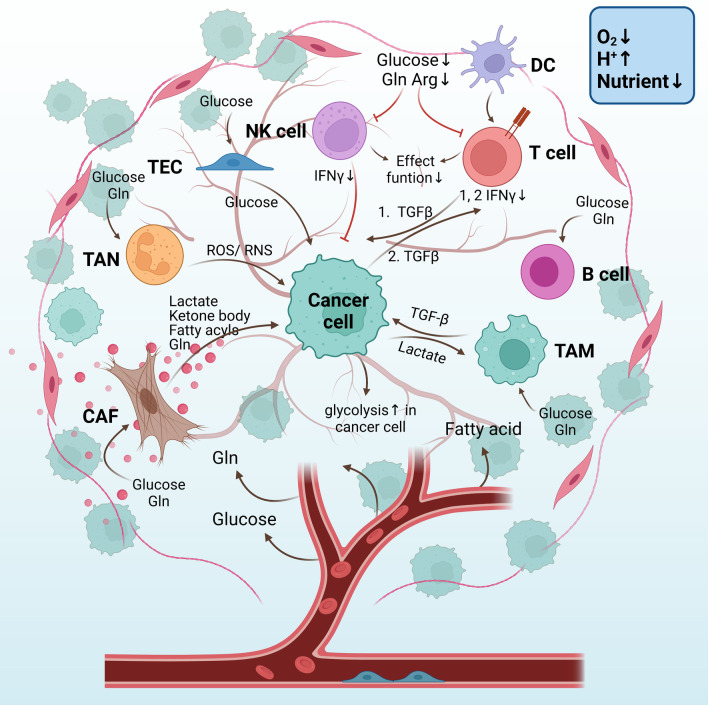
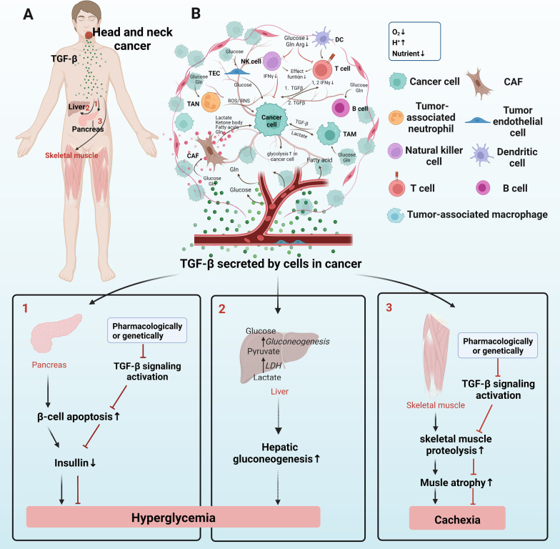
Transforming growth factor-β (TGF-β) signaling has a paradoxical role in cancer progression, and it acts as a tumor suppressor in the early stages but a tumor promoter in the late stages of cancer. Once cancer cells are generated, TGF-β signaling is responsible for the orchestration of the immunosuppressive tumor microenvironment (TME) and supports cancer growth, invasion, metastasis, recurrence, and therapy resistance. These progressive behaviors are driven by an "engine" of the metabolic reprogramming in cancer. Recent studies have revealed that TGF-β signaling regulates cancer metabolic reprogramming and is a metabolic driver in the tumor metabolic microenvironment (TMME). Intriguingly, TGF-β ligands act as an "endocrine" cytokine and influence host metabolism. Therefore, having insight into the role of TGF-β signaling in the TMME is instrumental for acknowledging its wide range of effects and designing new cancer treatment strategies. Herein, we try to illustrate the concise definition of TMME based on the published literature. Then, we review the metabolic reprogramming in the TMME and elaborate on the contribution of TGF-β to metabolic rewiring at the cellular (intracellular), tissular (intercellular), and organismal (cancer-host) levels. Furthermore, we propose three potential applications of targeting TGF-β-dependent mechanism reprogramming, paving the way for TGF-β-related antitumor therapy from the perspective of metabolism.

Keywords: Cancer cell; Host metabolism; Stromal cell; TGF-β signaling; Tumor metabolic microenvironment.


Conflict of interest statement

All other authors declare no competing interests.

Figures

Fig. 1
Fig. 1
Schematic representation of the gene TGFB(A) and protein TGF-β. A Gene structure of TGFB1, TGFB2, and TGFB3: The blue boxes represent the exons; the 5’- and 3’-untranslated region are marked in pink and green boxes, respectively. B Latent TGF-β synthesis and secretion: TGF-β precursor protein consists of a signal peptide, a LAP prodomain, and a mature TGF-β monomer sequence. With the removal of signal peptide, the precursor proteins are dimerized. After proteolytic cleavage, the mature TGF-β dimer remains associated with LAP prodomains and the SLC is formed. Then, SLC links with LTBP or LRRC and thus LLC is generated. The LLC is then secreted into extracellular matrix. C, D Once released from cells, the TGF-β dimer that is kept inactive by its binding with LTBP, which targets latent TGF-β into the ECM, or with an LRRC molecule that fixes latent TGF-β at the surface of cells. D, E integrin β, in association with integrin α, can bind with the RGD sequence in the latent TGF-β complex. Then, the increased tension at the interface leads to degradation of the LAP, and the physiological activation of latent TGF-β complexes result in the release of TGF-β ligands. These active TGF-β ligands bind to the TGFβRI/TGFβRII receptor complex at the cell surface, and the intracellular TGF-β signaling is initiated. LAP: latency-associated polypeptide or LAP. SLC: small latent complex; LTBP: latent TGF-β-binding protein; LLC: large latent complex; LRRC: leucine-rich repeat containing; RGD: arginine–glycine–aspartic acid motif. The short solid lines represent covalent bonds, while the short dashed lines are non-covalent bonds
Fig. 2
Fig. 2
Canonical and non-canonical TGF-β signaling pathway. LLC are produced by various cell types, and TGF-β ligands can be activated and release from the LLC. TGF-β signaling initiates when the TGF-β ligands (TGF-β1,2,3) bind to TGF-βRII.  Then, TGF-βRI is phosphorylated and activates various intracellular signaling cascades. These intracellular pathways of canonical SMAD2/3 pathway and non-canonical TGF-β pathways including MAP kinases (p38), Hippo, PI3K/AKT, and AMPK signaling can subsequently regulate metabolic activities. LLC: large latent complex
Fig. 3
Fig. 3
TGF-β-dependent metabolism reprogramming of cancer cells. 1. For glucose metabolism, TGF-β signaling can: a. enhance glycolysis by directly increasing the glycolytic enzyme expression of PKM2 and LDH, and indirectly elevate the PFKFB3, which augments the expression of PFK1, one of the glycolytic enzymes; b. TCA cycle enzyme SDH mutation leads to TGF-β-induced EMT; c. augment PPP by increasing G6PD, the first key enzyme of PPP; and d. promote glycogen synthesis by boosting GYS1 expression via inhibiting GSK-3 that deactivates GYS1. 2. For cholesterol metabolism, a. cholesterol-enriched subdomain-mediated TGF-β receptor endocytosis can: destruct TGF-β receptors that abrogate TGF-β signaling initiation; b. cholesterol synthesis enzyme NSDHL can inhibit TGF-β1 production and attenuate TGF-β signaling; c. NSDHL also prevents TGF-βRII endocytosis and then facilitates TGF-β signaling; and d. TGF-β signaling can decrease cholesterol synthesis by inactivating SREBF2, a cholesterol synthesis promoter. 3. For fatty acid metabolism, a. canonical (p-SMAD2) and non-canonical (p-AMPK) TGF-β signaling accelerates FAO; b. TGF-β1 can increase FASN expression, thus allowing fatty acid synthesis and accumulation in cancer cells; conversely, FASN enhances TGF-β1 production. Moreover, non-canonical (p-AMPK) signaling decreases FASN; c. TGF-β2 releasement by the acidic TMME enables more fatty acid entry into cancer cells and forms LD for lipid storage used by FAO. 4. For amino acid metabolism, TGF-β signaling can: a. increase Asp, Glu, and Lys, while decrease Ala, Asn, Gln, etc.; b. bolster glutamine’s entry into cancer cells by elevating SLC7A5, and enhance glutamine’s catalyzation into glutamate via increasing GLS1, and glutamate can flow into TCA cycle. Green highlighted items mean TGF-β signaling positively regulates them, or they are positively regulated by TGF-β signaling. Red vice versa. And these green and red ones are potentially TGF-β-dependent metabolic targets in cancer. HK2: Hexokinase 2; G6P: glucose 6-phosphate; F6P: fructose 6-phosphate; PFK1: phosphofructokinase 1; PFKFB3: 6-phosphofructo-2-kinase/fructose-2,6-biphosphatase 3; PPP: pentose phosphate pathway; Acetyl-CoA: acetyl coenzyme A; GYS1: glycogen synthase 1; GSK-3: glycogen synthase kinase 3; F-1,6-P: fructose-1,6-bisphosphate; G3P: glyceraldehyde 3-phosphate; PEP: phosphoenolpyruvate; PKM2: pyruvate kinase M2; LDH: Lactate dehydrogenase; SREBF2: sterol regulatory element-binding transcription factor 2; FASN: fatty acid synthase; FAO: fatty acid oxidation; LD: lipid droplet. ASP: aspartic acid; Glu: glutamic acid; Lys: lysine; Ala: alanine; Asn: asparagine; Gln: glutamine; Gly: glycine; His: histidine; Ile: isoleucine; Leu: leucine; Phe: phenylalanine; Pro: proline; Thr: threonine; Tyr: tyrosine. SLC7A5: glutamine transporter solute carrier family 7 member 5; and GLS1: glutaminase 1
Fig. 4
Fig. 4
TGF-β-dependent CAF–cancer cell metabolic coupling. 1. TGF-β signaling increases RWE of CAFs via decreasing CAV-1 and IDH3α, while increasing PDK1, PKM2, and ROS. ROS conversely promotes TGF-β signaling, which sustains CAF phenotype. In this way, aerobic glycolysis of CAFs increases and a large amount of lactate molecules are produced. Lactate shuttles from CAFs to cancer cells via MCT-4 and MCT-1. Lactates are then converted to pyruvate and utilized for TCA cycle of cancer cell. 2. Glutamine, ketone body, and BCKAs are also substrates of RWE that are produced by CAFs and then are transferred into cancer cells for TCA cycle of cancer cell. 3. TGF-β-mediated metabolic coupling can also correlate with glycogen metabolism. Cancer cell-derived TGF-β cytokines trigger the TGF-β non-canonical p38-MAPK signaling in CAFs via paracrine, which stimulates the production of several cytokines including IL-6, CXCL10, and CCL5 from CAFs. These cytokines induce glycogen metabolism upregulation in cancer cells via phosphorylation and activation of PGM1, an enzyme that is involved in glycogen synthesis. Then glycogen is accumulated in cancer cells and promotes cancer cell metastasis. Green highlighted items mean TGF-β signaling positively regulates them or they are positively regulated by TGF-β signaling. Red vice versa. And these green and red ones are potentially TGF-β-dependent metabolic targets in cancer. PDK1: pyruvate dehydrogenase kinase 1; ROS: reactive oxygen species; RWE: reverse Warburg effect; BCAAs: branched-chain amino acids; BCKAs: branched-chain α-ketoacids. BCAT1: BCAA transaminases; and PGM1: phosphoglucomutase 1
Fig. 5
Fig. 5
The “fruit tree” schematic diagram of the metabolic coupling between stromal and cancer cells through nutrients and metabolites. CAFs provide mitochondrial fuels for cancer cells, including lactate, ketone body, fatty acyls, and Gln. TANs can release ROS and RNS to kill cancer cells directly. TECs absorb glucose molecules and then provide them to cancer cells. TAMs are influenced by lactate that is exported from cancer cells, while TAMs provide cancer cells with TGF-β which elevated the glycolytic rate of cancer cells. Malignant B cells tend to be glycolytic and Gln-addicted. Since all cells compete for nutrients of glucose, fatty acid, and amino acid, under this nutrient-deficient TMME, T cells and NK cells are immunosuppressive with less IFNγ releasement. TGF-β-mediated metabolic coupling with cancer cells mainly occurs in T cells based on previous literature. 1. Cancer cells with increased aerobic glycolysis by HK2 expression are easier to “educate” antitumor CD4 + T cell to be immune-suppressed through ways below: Stimulate CD4 + T cell secret TGF-β, an immune-suppressing cytokine; and glucose competition between cancer cells and CD4 + T cells lowers glycolytic rate in CD4 + T cells, which decreases the antitumor activity of CD4 + T cells by IFNγ production decline. 2. Cancer cell-derived TGF-β can also decrease INFγ secretion of CD4+ T cells. Few/abnormal blood vessels lead to a hypoxic TMME(O2↓) which stimulates aerobic glycolysis in cancer cells and stromal cells. Lactate produced by glycolysis contributes to an acidic TMME(H+↑). Since all cells in TMME compete for the restricted nutrient, therefore nutrient is deficient (Nutrient↓) in TMME. CAFs: cancer-associated fibroblasts, Gln: glutamine; Arg: arginine; TAN: tumor-associated neutrophils; ROS: reactive oxygen species; RNS: reactive nitrogen species. TECs: tumor endothelial cells; TAMs: tumor-associated macrophages; Arg: arginine; TMME: tumor metabolic microenvironment; and HK2: Hexokinase 2
Fig. 6
Fig. 6
TGF-β-mediated host metabolism reprogramming in cancer. A Cancer is like a malignant organ that produces a large amount of TGF-β cytokine, then it circulates all over the host body including the liver, pancreas, and skeletal muscle. B In this way, TGF-β ligand resembles an endocrine factor and leads to hyperglycemia through 1. inducing β-cell apoptosis (verified) and 2. hepatic gluconeogenesis (hypothesized). 3. Cachexia is induced by TGF-β signaling by skeletal muscle proteolysis (verified). Pharmacologically or genetically inhibiting TGF-β signaling can attenuate these processes and reverse hyperglycemia and cachexia.

References

    1. Yeh HW, Lee SS, Chang CY, Lang YD, Jou YS. A new switch for TGFβ in cancer. Cancer Res. 2019;79(15):3797–3805. doi: 10.1158/0008-5472.CAN-18-2019. - DOI - PubMed
    1. Davis MD, Suzaki I, Kawano S, Komiya K, Cai Q, Oh Y, Rubin BK. Tissue factor facilitates wound healing in human airway epithelial cells. Chest. 2019;155(3):534–539. doi: 10.1016/j.chest.2018.10.007. - DOI - PMC - PubMed
    1. Hinck AP, Mueller TD, Springer TA. Structural biology and evolution of the TGF-β family. Cold Spring Harb Perspect Biol. 2016;8(12):a022103. doi: 10.1101/cshperspect.a022103. - DOI - PMC - PubMed
    1. Wynn TA, Barron L. Macrophages: master regulators of inflammation and fibrosis. Semin Liver Dis. 2010;30(3):245–257. doi: 10.1055/s-0030-1255354. - DOI - PMC - PubMed
    1. Batlle E, Massagué J. Transforming growth factor-β signaling in immunity and cancer. Immunity. 2019;50(4):924–940. doi: 10.1016/j.immuni.2019.03.024. - DOI - PMC - PubMed

Publication types