Review
. 2022 Nov:351:504-559.
doi: 10.1016/j.jconrel.2022.09.024. Epub 2022 Sep 30.

Mucoadhesive carriers for oral drug delivery

Affiliations
Review

Mucoadhesive carriers for oral drug delivery

Raj Kumar et al. J Control Release. 2022 Nov.

Abstract

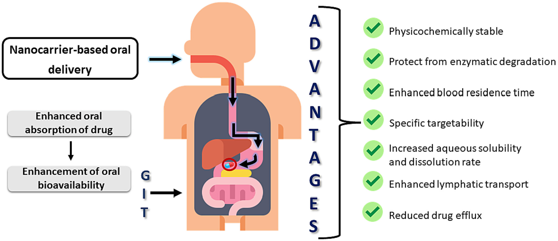
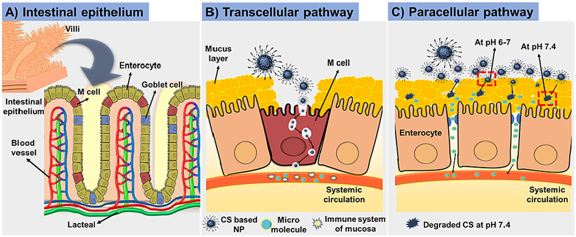
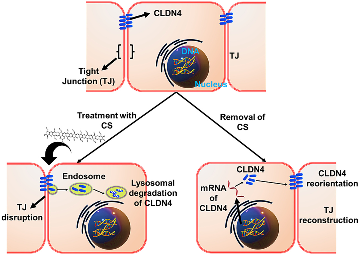
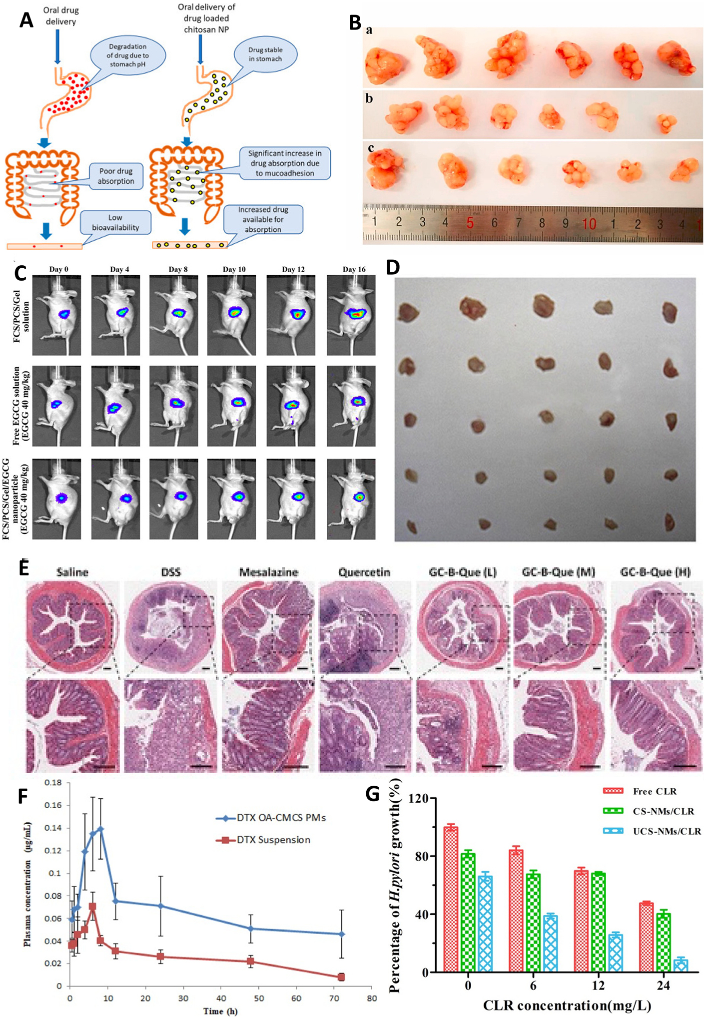
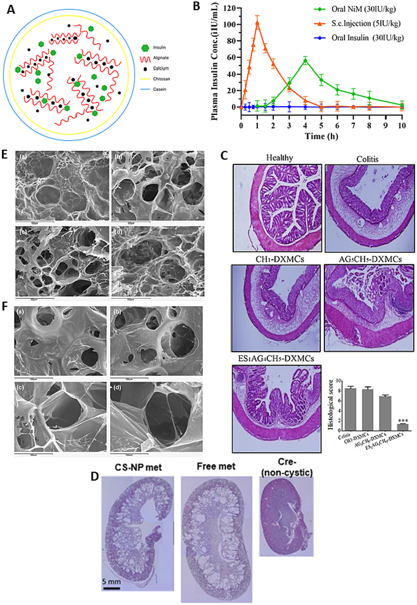
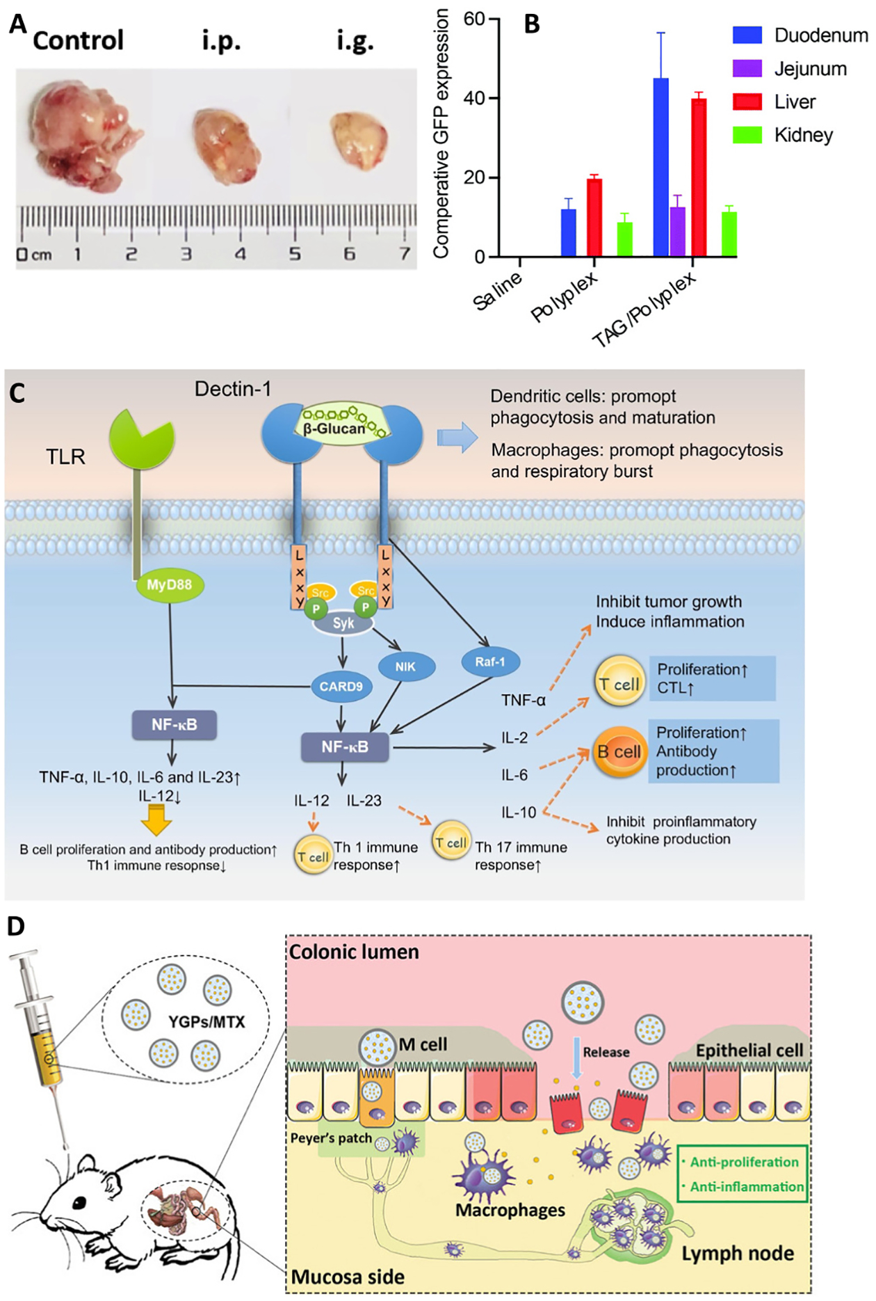
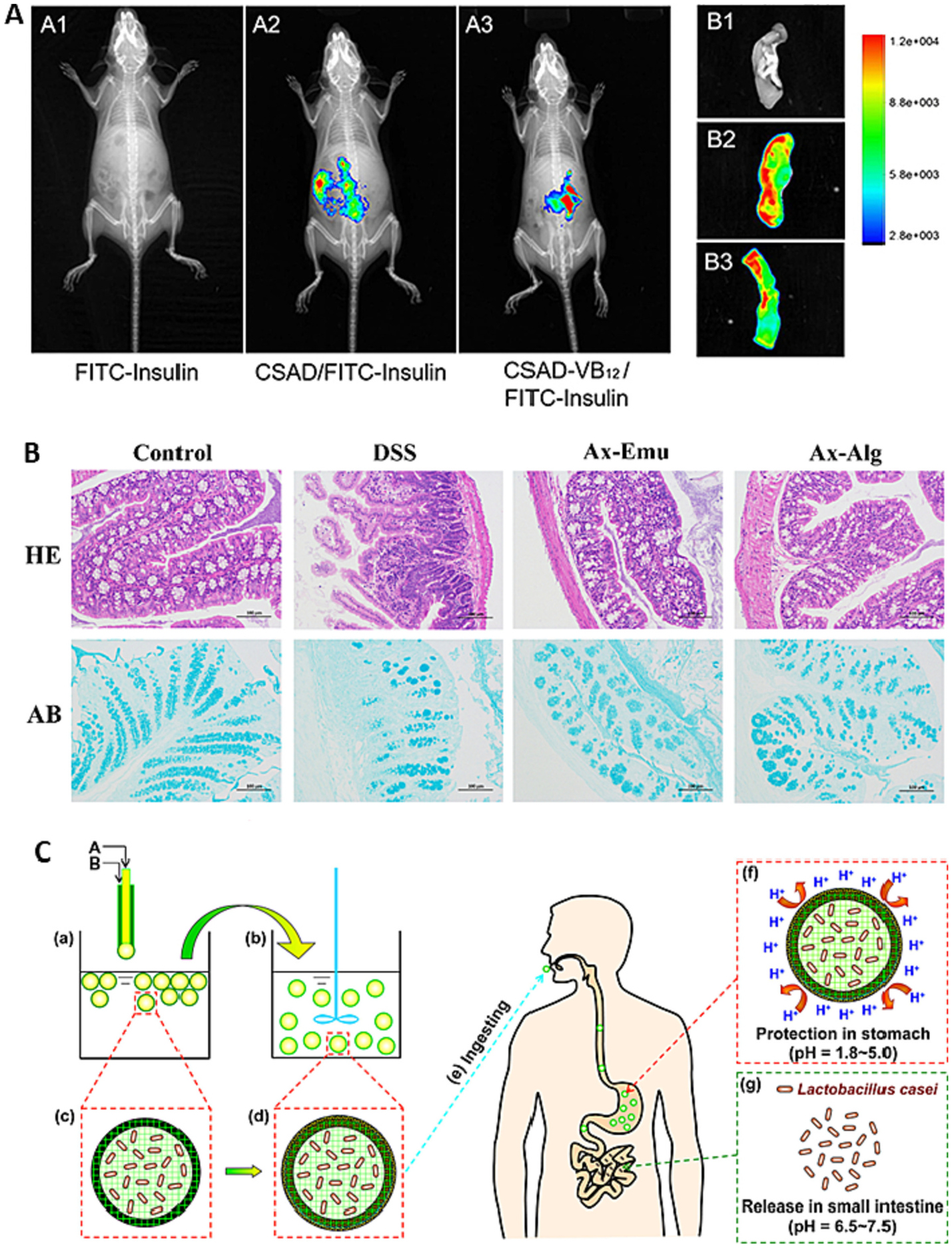
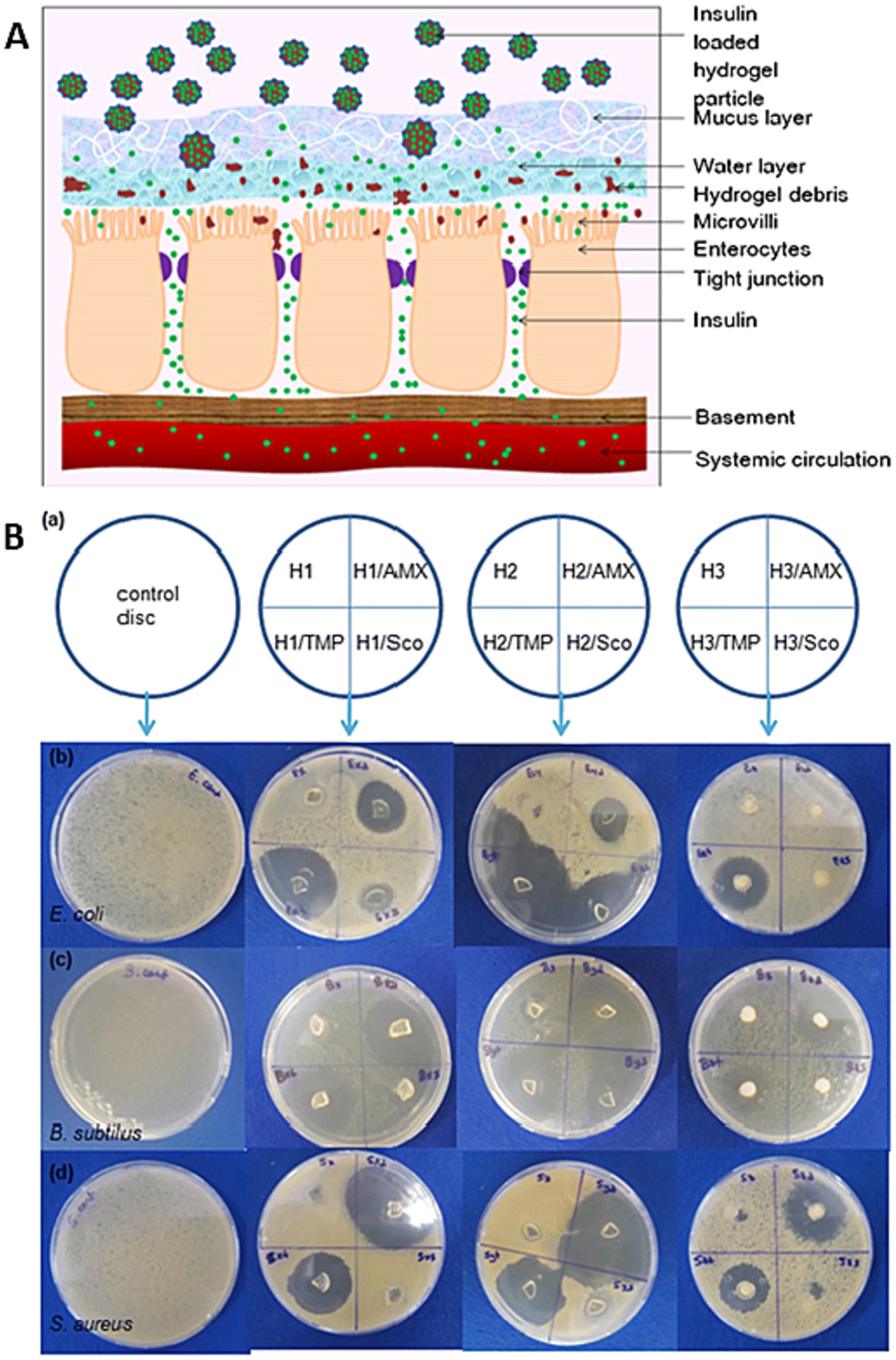
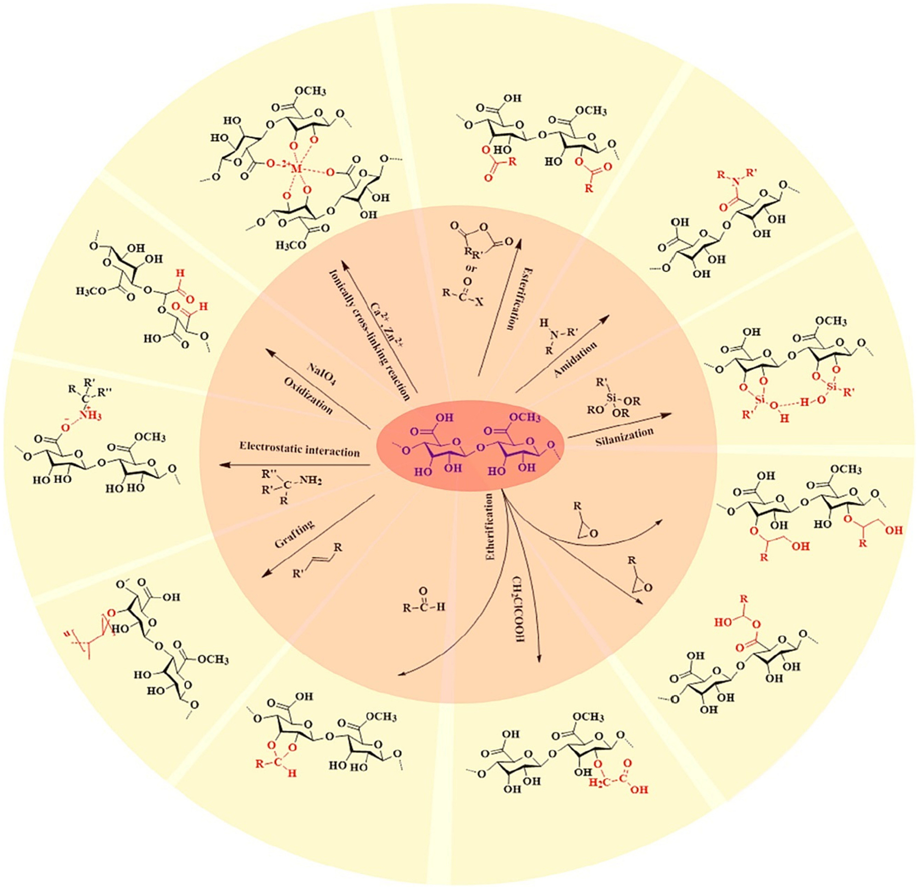
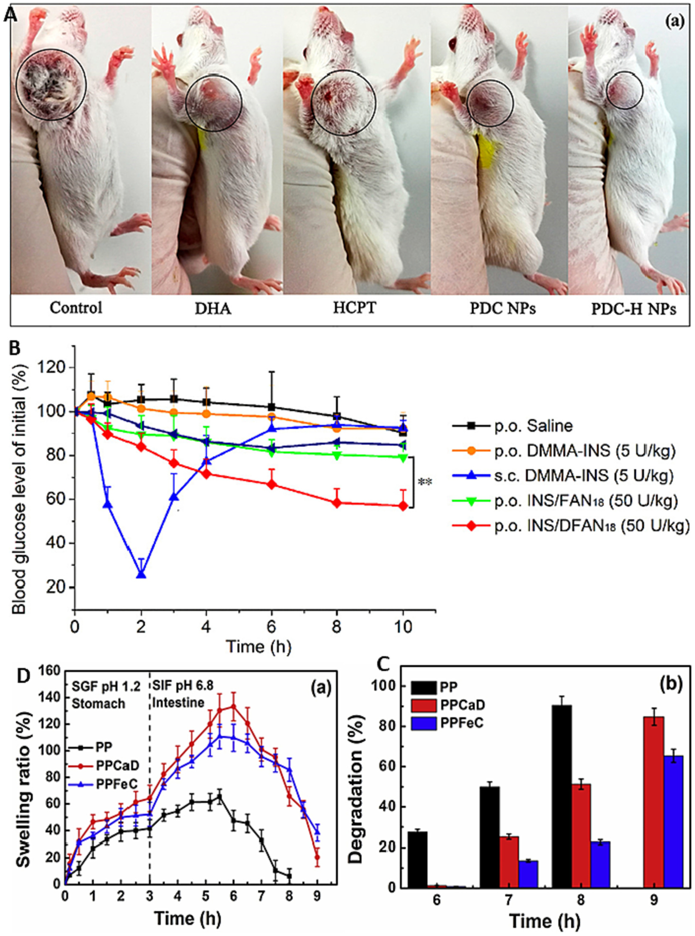
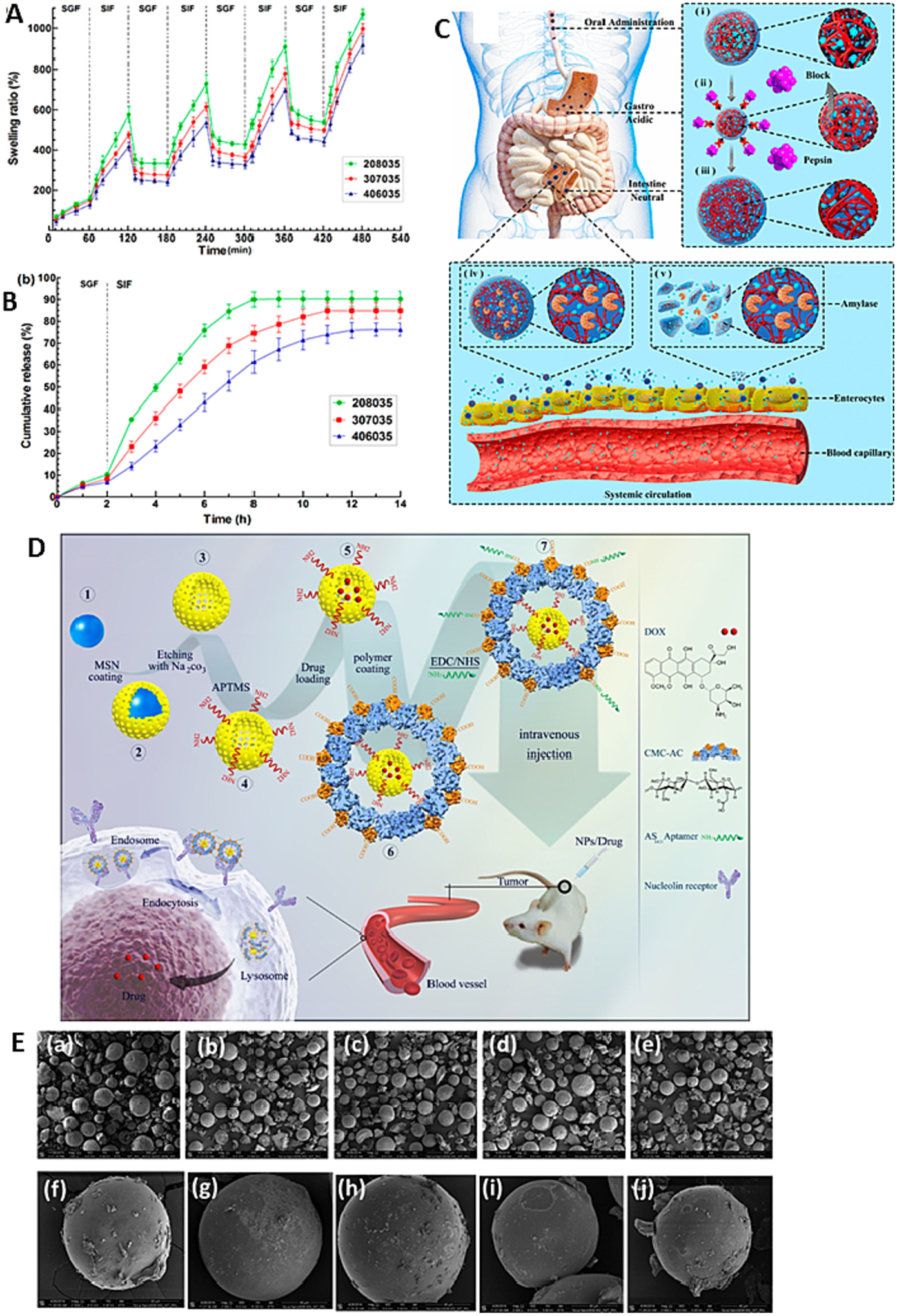
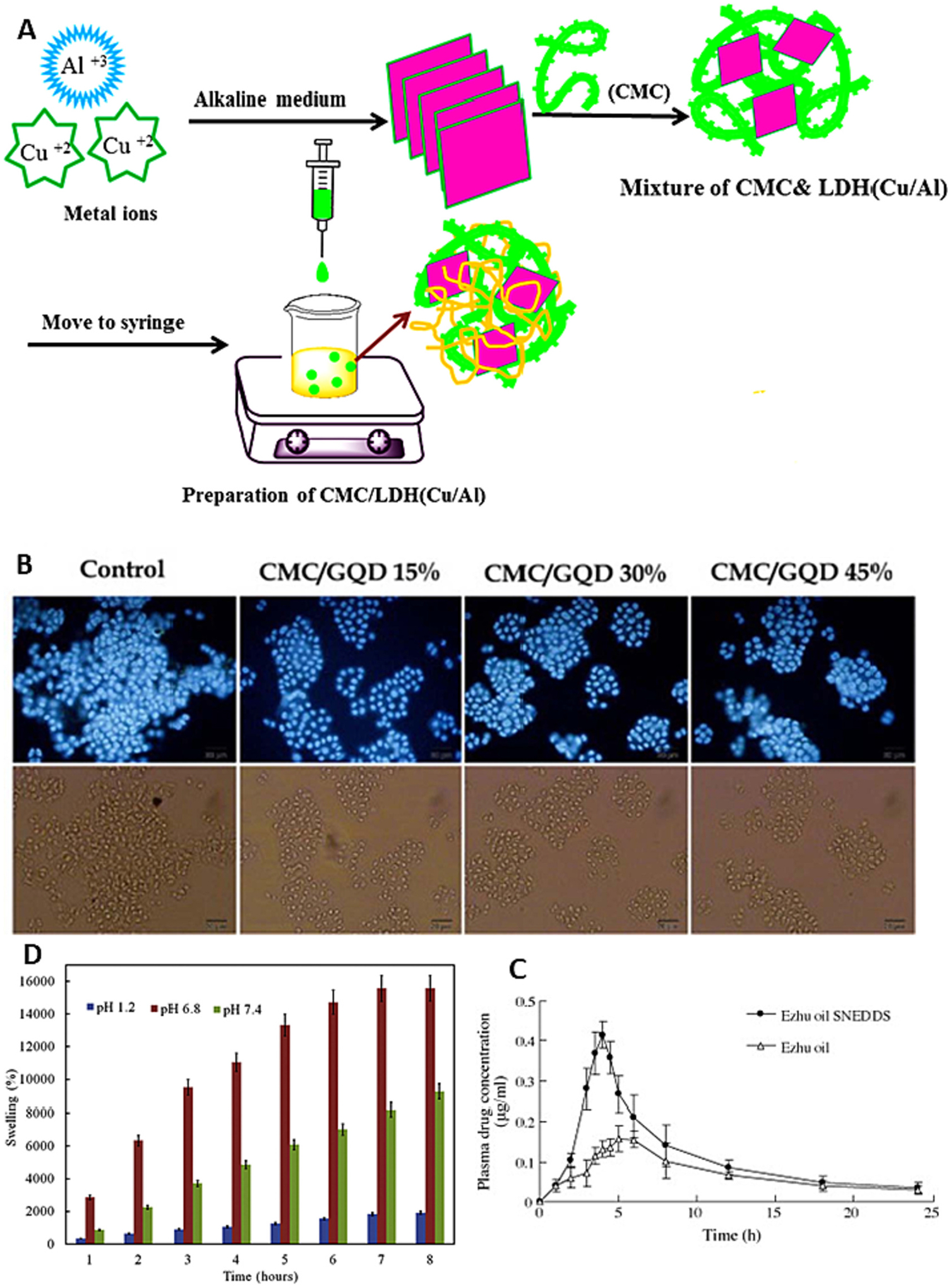
Among the various dosage forms, oral medicine has extensive benefits including ease of administration and patients' compliance, over injectable, suppositories, ocular and nasal. Despite of extensive demand and emerging advantages, over 50% of therapeutic molecules are not available in oral form due to their physicochemical properties. More importantly, most of the biologics, proteins, peptide, and large molecular drugs are mostly available in injectable form. Conventional oral drug delivery system has limitation such as degradation and lack of stability within stomach due to presence of highly acidic gastric fluid, hinders their therapeutic efficacy and demand more frequent and higher dosing. Hence, formulation for controlled, sustained, and targeted drug delivery, need to be designed with feasibility to target the specific region of gastrointestinal (GI) tract such as stomach, small intestine, intestine lymphatic, and colon is challenging. Among various oral delivery approaches, mucoadhesive vehicles are promising and has potential for improving oral drug retention and controlled absorption to treat local diseases within the GI tract, as well systemic diseases. This review provides the overview about the challenges and opportunities to design mucoadhesive formulation for oral delivery of therapeutics in a way to target the specific region of the GI tract. Finally, we have concluded with future perspective and potential of mucoadhesive formulations for oral local and systemic delivery.

Keywords: Gastric cancer; Inflammatory bowel disease; Mucoadhesive polymer; Oral delivery; Oral medicine.


Figures

Fig. 1.
Fig. 1.
The scheme represents potential benefits of mucoadhesive-based formulation for oral drug delivery.
Fig. 2.
Fig. 2.
Schematic illustrations of (A) the structure of the intestinal epithelium (B) the transcellular and (C) paracellular transport of nanoparticles.
Fig. 3.
Fig. 3.
Schematic mechanism of chitosan mediated reversible tight junction (TJ) opening.
Fig. 4.
Fig. 4.
(A) in vivo efficiency of chitosan nanoparticles increasing absorption of drug through intestinal epithelium; (B) Photos of original tumor size. (a) control, (b) COS–Se (100 mg/kg), and (c) COS-Se (50 mg/kg); (C) The antitumor activities using a noninvasive in vivo imaging of Orthotopic Luc MKN45 xenograft models treated with different sample daily; (D) In vivo antitumor efficacy study (E) in vivo anti-inflammatory activity of smart responsive quercetin-conjugated glycol chitosan prodrug micelles, Histological study of inflammatory tissues after treatment by hematoxylin and eosin staining; (F) Plasma concentration vs time profile of DTX (Docetaxel) after oral administration of 10 mg/kg dose of DTX suspension and DTX loaded OA-CMCS micelles; (G) In vitro anti-bacterial activity of free clarithromycin (CLR), clarithromycin-loaded CMCS-g-SA nano-micelles (CS-NMs/CLR) and clarithromycin-loaded U-CMCS-g-SA nano-micelles (UCS-NMs/CLR) against H. pylori;. Images of tumor tissues. Reproduced with permission from ref. [–98]. Copyright 2019 Elsevier, 2017 MDPI open access, 2020 Elsevier, 2015 American Chemical Society, 2021 American Chemical Society, 2020 Elsevier, 2019 Elsevier.
Fig. 5.
Fig. 5.
(A) Schematic representation of NiM particles; (B) Plasma insulin concentration-time curves after subcutaneous injection of insulin solution (5 IU/kg), oral administration of NiM (30 IU/kg), and oral administration of insulin solution (30 IU/kg) in diabetic mice; (C) Histological assessment of colitis in colon tissues from the different study groups shown remarkable in histological feature in mice treated with ES1AG4CH5-DXMCs; (D) H&E staining of whole kidneys shows less severe cystic phenotype in the CS-NP met group; (E) Scanning electron micrographs of CAM:[CS:CMC] 40:[25:75] (w/w) complexes. The first two micrographs depict top view of mini matrices at 30 min and pH 1.2 with low M.wt. CS (a) and high M.wt. CS (b). The other two micrographs outline surfaces at 1 h and pH 1.2: low M.wt. CS (c) and high M.wt. CS (d); (F) Scanning electron micrographs of CAM: [CS: CMC] 40:[25:75] (w/w) complexes. The first two micrographs depict top view of mini matrices at 1 h and pH 4.2 with low M.wt. CS (a) and high M.wt. CS (b). The other two micrographs outline surfaces at 4 h and pH 4.2: low M.wt. CS (c) and high M. wt. CS (d). reproduced with permission from ref. [,–136]. Copyright 2008, 2020, 2018 Elsevier, and 2021 Wiley.
Fig. 6.
Fig. 6.
(A) representative photographs of S-180 tumors of mice were subcutaneously inoculated with S-80 cells. 2 mg/kg AG was intraperitoneally or orally administrated to mice daily after tumor inoculation for 1 week: (B) Quantitative expression of eGFP in different tissues/organs of orally administered polyplex and TAG/polyplex. TAG containing particles show duodenumspecific transportation and liver accumulation. Liver accumulation of eGFP for TAG-mediated particles was 3× higher than polyplex; (C) The signal pathway of β-glucan binding to dectin-1. Once they are bond, dectin-1 will activate Syk via double “LxxY” structures, and then trigger NF-jB mainly throughCARD9 or NIK to produce IL-10, IL-2, IL-23, IL-6 and TNF, resulting in T and B cells proliferation, DCs maturation and respiratory burst. Dectin-1 can also trigger NF-jB by Raf-pathway directly. When dectin-1 and MyD88 of TLRs are both activated, there will appear a coupling-signal that prompts the production of TNF-a, IL-10, IL-6 and IL-23, and down regulates the expression of IL-12; (D) The preparation process of YGPs/MTX and the illustration of YGPs/MTX targeting inflammatory sites and suppressing intestinal inflammation after intragastric administration. Reproduced with permission from ref. [,–182]. Copyright 2019 Elsevier, 2022 The Royal Society of Chemistry, 2018 Elsevier, and 2020 Wiley-Vch.
Fig. 7.
Fig. 7.
(A) The retention effect and absorption of FITC-insulin, CSAD/FITC-insulin, CSAD-VB12/FITC-insulin in intestine. (A) In vivo animal image system showed the fluorescent signal of FITC-insulin (A1), CSAD/FITC-insulin (A2), CSAD- VB12/FITC-insulin (A3) at the 1 h after oral administration in T1D mice. (B) Representative fluorescence imaging of small intestine from mice treated with FITC-insulin (B1), CSAD/FITC-insulin (B2), CSAD- VB12/FITC-insulin (B3); (B) Histological score of colon tissues by hematoxylin and eosin (HE) and alcian blue (AB) staining after treatment of Ax-Alg to DSS colitis mice. (C) Schematic illustration of the preparation process and the design concept of the proposed intestinal-targeted CAP carrier for pH-responsive protection and release of lactic acid bacteria. (a, c) CA beads prepared by a coextrusion method. “A” is Na-alginate solution containing Lactobacillus casei, and “B” is pure Na-alginate solution. (b, d) CAP beads prepared by adsorption of protamine molecules. (e) Ingesting CAP beads in mouth. (f) CAP beads offer improved protection for Lactobacillus in stomach. (g) CAP beads release Lactobacillus casei rapidly in the small intestine. Reproduced with permission from ref. [–201]. Copyright 2022 Elsevier, 2019 Dove press, open access, and 2014 American Chemical Society.
Fig. 8.
Fig. 8.
(A) Schematic diagram showing the release and absorption of insulin from intestine following oral administration of hydrogel-based systems; (B) Digital photographs of antimicrobial activity tests of drug-loaded and unloaded semiIPN hydrogels (a) Schematic representation of the test application for control disc, H1, H2 and H3 hydrogels (top to bottom), or the bacteria applied (b) E. coli. (c) B. subtilus (d) S. aureus. Reproduced with permission from ref. [218,220,220]. Copyright 2012 Elsevier, 2020 the Polymer Society, Taipei.
Fig. 9.
Fig. 9.
Potential reactions of pectin for synthesis of various derivatives. Reproduced with permission from the ref [243]. Copyright 2021 Elsevier.
Fig. 10.
Fig. 10.
(A) Tumor photographs of each treatment group on day 24 of In vivo antitumor activity of free DHA, free HCPT, and nanoparticles in the subcutaneous mouse model of 4T1 cells. Tumor photographs of each treatment group on day 24; (B) Blood glucose level vs. time profiles of STZ-induced diabetic rats administrated with insulin solution, INS/FAN18 and INS/DFAN18 through different routes; (C) (a) Swelling ratio of the hydrogels PP, PPCaD and PPFeC in SGF at pH 1.2 and in SIF at pH 6.8 (b) degradation behavior in SIF at pH 6.8 for various hydrogels. Reproduced with permission from ref. [–249]. Copyright 2022, 2019 Elsevier, and 2017 American Chemical Society.
Fig. 11.
Fig. 11.
(A) Dynamic swelling/deswelling of hydrogels in simulated gastric fluid (SGF) and simulated intestinal fluid (SIF); (B) in vitro BSA release profile of the hydrogels in SGF and SIF; (C) Proposed Schematic Illustration for the Behaviors of the Insulin-Loaded Microgels during the Delivery Process in the GI Tract; (D) Schematic presentation of the preparation of HMSNs, their coating with acetylated CMC and functionalization with AS1411 aptamer for targeting nucleolin over-expressing cancer cells; (E) SEM Micrographs of (a) MS1, (b) MS2, (c) MS3, (d) MS4, (e) MS5 microspheres at 150 magnification and single (f) MS1, (g) MS2, (h) MS3, (i) MS4, (j) MS5 microspheres at 2000 magnification. Reproduced with permission from ref. [265,286,290,291]. Copyright 2014 American Chemical Society, 2018 Elsevier, 2014 The Royal Society of chemistry, 2018 American Chemical Society.
Fig. 12.
Fig. 12.
(A) Schematic procedure for preparation of CMC/LDH(Cu/Al) bio-nanocomposite hydrogel bead; (B) DAPI stained nuclei of colon cancer cells (HT29 cells) with CMC/GQDs at various GQDs percentage (15%, 30%, and 45%); (C) Plasma concentration of GM after oral administration of unformulated ZTO (500 mg/kg) or ZTO-SNEDDS (1625 mg/kg) to SD rats; (D) Swelling behavior of CMC/Cu-MOF@IBU at pH values of 1.2, 6.8 and 7.4. Reproduced with permission from ref. [315,317,320,321]. Copyright 2019, 2019, 2009, 2018 Elsevier.

References

    1. Agrawal U, Sharma R, Gupta M, Vyas SP, Is nanotechnology a boon for oral drug delivery? Drug Discov. Today 19 (2014) 1530–1546, 10.1016/J.DRUDIS.2014.04.011. - DOI - PubMed
    1. Ensign LM, Cone R, Hanes J, Oral drug delivery with polymeric nanoparticles: The gastrointestinal mucus barriers, Adv. Drug Deliv. Rev 64 (2012) 557–570, 10.1016/J.ADDR.2011.12.009. - DOI - PMC - PubMed
    1. Banerjee A, Qi J, Gogoi R, Wong J, Mitragotri S, Role of nanoparticle size, shape and surface chemistry in oral drug delivery, J. Control. Release 238 (2016) 176–185, 10.1016/J.JCONREL.2016.07.051. - DOI - PMC - PubMed
    1. Mei L, Zhang Z, Zhao L, Huang L, Yang XL, Tang J, Feng SS, Pharmaceutical nanotechnology for oral delivery of anticancer drugs, Adv. Drug Deliv. Rev 65 (2013) 880–890, 10.1016/J.ADDR.2012.11.005. - DOI - PubMed
    1. Bernkop-Schnürch A, Mucoadhesive systems in oral drug delivery, Drug Discov. Today Technol 2 (2005) 83–87, 10.1016/J.DDTEC.2005.05.001. - DOI - PubMed

Publication types