. 2022 Sep 19;20(1):298.
doi: 10.1186/s12957-022-02764-2.

The clinicopathological and prognostic factors of hepatocellular carcinoma: a 10-year tertiary center experience in Egypt

Affiliations

The clinicopathological and prognostic factors of hepatocellular carcinoma: a 10-year tertiary center experience in Egypt

Dina Sweed et al. World J Surg Oncol. .

Abstract

Background: Hepatocellular carcinoma (HCC) remains a major health problem despite the emergence of several preventive and therapeutic modalities. HCC has heterogeneous and wide morpho-molecular patterns, resulting in unique clinical and prognostic criteria. Therefore, we aimed to study the clinical and pathological criteria of HCC to update the morpho-molecular classifications and provide a guide to the diagnosis of this disease.

Methods: Five hundred thirty pathologically analyzed HCC cases were included in this study. The clinical and survival data of these cases were collected.

Results: Hepatitis C virus is still the dominant cause of HCC in Egypt. Post-direct-acting antiviral agent HCC showed an aggressive course compared to interferon-related HCC. Old age, male gender, elevated alpha-fetoprotein level, tumor size, and background liver were important prognostic parameters. Special HCC variants have characteristic clinical, laboratory, radiological, prognostic, and survival data. Tumor-infiltrating lymphocytes rather than neutrophil-rich HCC have an excellent prognosis.

Conclusions: HCC is a heterogenous tumor with diverse clinical, pathological, and prognostic parameters. Incorporating the clinicopathological profile per specific subtype is essential in the treatment decision of patients with HCC.

Trial registration: This was a retrospective study that included 530 HCC cases eligible for analysis. The cases were obtained from the archives of the Pathology Department, during the period between January 2010 and December 2019. Clinical and survival data were collected from the patients' medical records after approval by the institutional review board (IRB No. 246/2021) of Liver National Institute, Menoufia University. The research followed the guidelines outlined in the Declaration of Helsinki and registered on ClinicalTrials.gov (NCT05047146).

Keywords: DAAs; Hepatitis C virus; Hepatocellular carcinoma; Pathological subtypes; prognosis.


Conflict of interest statement

The authors declare that they have no competing interests.

Figures

Fig. 1
Fig. 1
The primary parameters used to conduct comparative analyses
Fig. 2
Fig. 2
Different macroscopic appearance of HCC. a Solitary pseudocapsulated nodule in cirrhotic liver. b A diffuse or cirrhometric HCC with multiple satellite nodules in non-cirrhotic liver. c An infiltrative HCC. d A solitary pseudocapsulated HCC; however, in non-cirrhotic liver. e A macroscopic bile duct invasion in HCC cases (blue boxes). f A macroscopic lymphovascular invasion in HCC case (blue box)
Fig. 3
Fig. 3
Microscopic aspects of HCC, NOS. a A well differentiated HCC showed thin trabaecule (IHC, 100×). b A moderately differentiated HCC showed wide acini filled with eosinophilic to bile secretion (IHC, 100×). c A moderately differentiated HCC showed mixed acinar and trabecular pattern (IHC, 100×). d A poorly differentiated HCC showed solid pattern with marked nuclear atypia (IHC, 100×). e HCC, NOS with mild intra-tumoral lymphocytes (IHC, 100×). f HCC, NOS with hemangiopericytoma like pattern (IHC, 100×). g HCC, NOS with prominent osteoclast like giant cells (IHC, 100×). h HCC with prominent lymphovascular invasion (arrows) (IHC, 100×). i HCC with bile duct invasion with attached tumor emboli to the epithelial cells (arrows) (IHC, 100×). j HCC with perineural invasion (arrows) (IHC, 100×). k HCC, NOS associated with calcified bilharzial ova (arrows) (IHC, 100×)
Fig. 4
Fig. 4
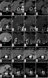
Contrast-enhanced triphasic CT imaging of typical and atypical HCC cases: A case of typical radiological appearance of multiple HCC (LR-5) (ad). a Cirrhotic liver changes, b with right hepatic lobe focal lesion seen at segment VI displaying intense arterial enhancement, c with washout of contrast in portovenous phase, d being hypo dense to hepatic parenchyma in delayed equilibrium study. eh Another focal lesion is seen at segment VII with similar enhancement pattern. A case of infiltrative HCC (LR-5) (il). i Cirrhotic liver changes, j with malignant infiltration of the left hepatic lobe that shows heterogeneous enhancement in the arterial phase with low density areas indicative of necrosis k and displays wash out of contrast at portovenous phase, l being hypodense to hepatic parenchyma in delayed equilibrium study. A case of multiple hypovascular HCC (LR-5) (mp). m Cirrhotic liver changes, np with multiple bilobar variable sized hepatic focal lesions showing no contrast uptake in different study phases, the largest at left hepatic lobe segment II measuring 5 × 4.8 cm. A case of HCC on top of non-cirrhotic liver (LR-M) (qt). q Non cirrhotic liver, r with right hepatic lobe segment VI large exophytic well defined focal mass lesion displaying thick irregular peripheral arterial enhancement and central hypo dense area of necrosis, s with washout of contrast at portovenous phase, t and delayed phases
Fig. 5
Fig. 5
Microscopic aspects of special HCC variants. a A macrotrabecular massive subtype showed trabeculae > 10 cells in thickness (IHC, 100×). b A clear cell subtype showed sheets of hepatocytes contained high glycogen and lipid content (IHC, 100×). c A clear cell subtype showed focal fatty changes (IHC, 100×). d A clear cell subtype showed severe fatty changes (IHC, 100×). e A steatohepatitic subtype showed a triad of fatty change, intra-tumoral fibrosis, and inflammation (IHC, 100×). f A steatohepatitic subtype showed Mallory hyaline bodies (IHC, 200×). g A scirrhous subtype showed compressed cords of hepatocytes within desmoplastic stroma (IHC, 100×). h A FLC variant showed hepatocytes with abundant eosinophilic cytoplasm, prominent eosinophilic nucleoli separated by lamellated collagen bundles (IHC, 100×). i A FLC showed intra-tumoral pale bodies (IHC, 200×)
Fig. 6
Fig. 6
Microscopic aspects of special HCC variants, continued. a A sarcomatoid subtype showed spindle tumor cells arranged in fascicular pattern (IHC, 100×). b A chromophobe subtype showed sheets of tumor cells with clear to eosinophilic cytoplasm, sharp cell border, and paranuclear halos (IHC, 100×). c A chromophobe subtype showed bland nuclei with area of abrupt anaplasia (IHC, 200×). d A LEL-HCC subtype showed intra-tumoral lymphocytes outnumbered the tumor cells (IHC, 100×). ef A neutrophil-rich subtype showed numerus and diffuse neutrophils within the tumor (IHC, 200×). g A c-HCC-CC showed mixed hepatocytic and cholangiocytic areas of differentiation (IHC, 100×). h c-HCC-CC showed the hepatocytic differentiation area (IHC, 100×). i c-HCC-CC showed the cholangiocytic differentiation area (IHC, 100×)
Fig. 7
Fig. 7
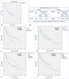
Survival data of HCC patients. a Kaplan-Meier survival curve demonstrating the OS of HCC patients. b Table demonstrated the mean and median survival time for HCC patients. c Kaplan-Meier survival curve demonstrating the impact of patients’ age on the OS. d Kaplan-Meier survival curve demonstrating the impact of tumor size on the OS. e Kaplan-Meier survival curve demonstrating the impact of TILs on the OS. f Kaplan-Meier survival curve demonstrating the impact of intra-tumoral fibrous stroma on the OS

Similar articles

Cited by

References

    1. Siegel RL, Miller KD, Fuchs HE, Jemal A. Cancer Statistics, 2021. CA Cancer J Clin. 2021;71(1):7–33. doi: 10.3322/caac.21654. - DOI - PubMed
    1. Holah N, El-Azab D, Aiad H, Sweed D. Hepatocellular carcinoma in Egypt: epidemiological and histopathological properties. Menoufia Med J. 2015;28(3):718–724.
    1. Blach S, Zeuzem S, Manns M, Altraif I, Duberg A-S, Muljono DH, et al. Global prevalence and genotype distribution of hepatitis C virus infection in 2015: a modelling study. Lancet Gastroenterol Hepatol. 2017;2(3):161–176. doi: 10.1016/S2468-1253(16)30181-9. - DOI - PubMed
    1. Rashed WM. Current HCC Clinical and Research in Egypt. In: Liver Cancer in the Middle East. Cham: Springer; 2021. p. 313–21.
    1. Wang X, Wang Z, Wu L. Combined measurements of tumor number and size helps estimate the outcome of resection of Barcelona clinic liver cancer stage B hepatocellular carcinoma. BMC Surg. 2016;16:22. doi: 10.1186/s12893-016-0135-4. - DOI - PMC - PubMed

Associated data