. 2022 Sep 18;12(1):15637.
doi: 10.1038/s41598-022-19520-x.

Osteogenesis in human periodontal ligament stem cell sheets is enhanced by the protease-activated receptor 1 (PAR1) in vivo

Affiliations

Osteogenesis in human periodontal ligament stem cell sheets is enhanced by the protease-activated receptor 1 (PAR1) in vivo

Tomaz Alves et al. Sci Rep. .

Abstract

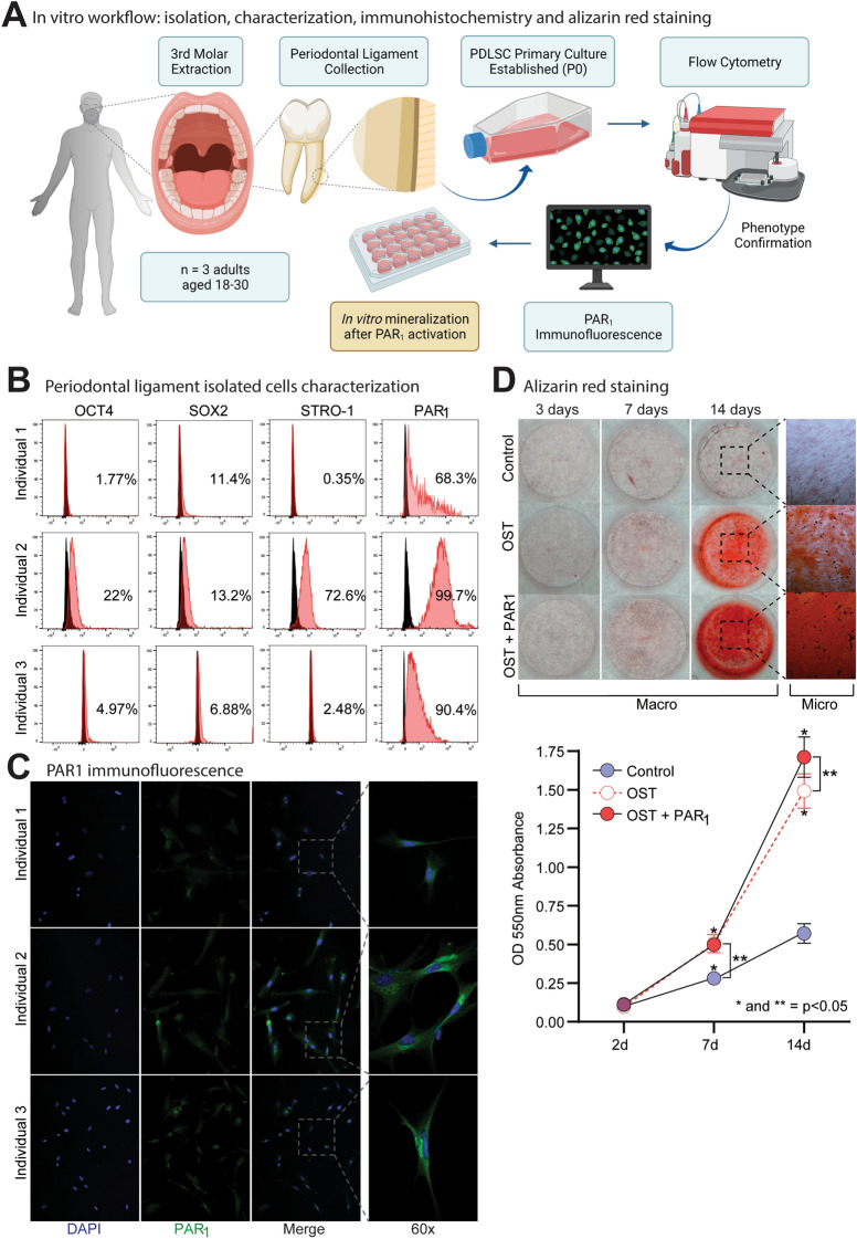
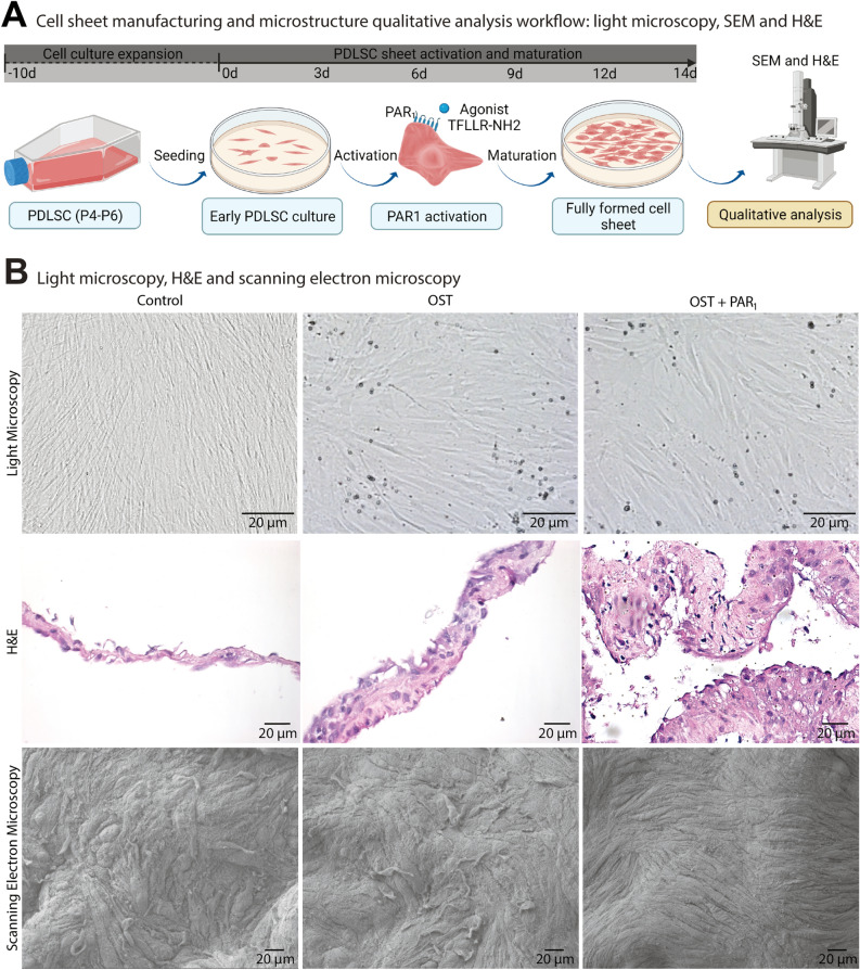
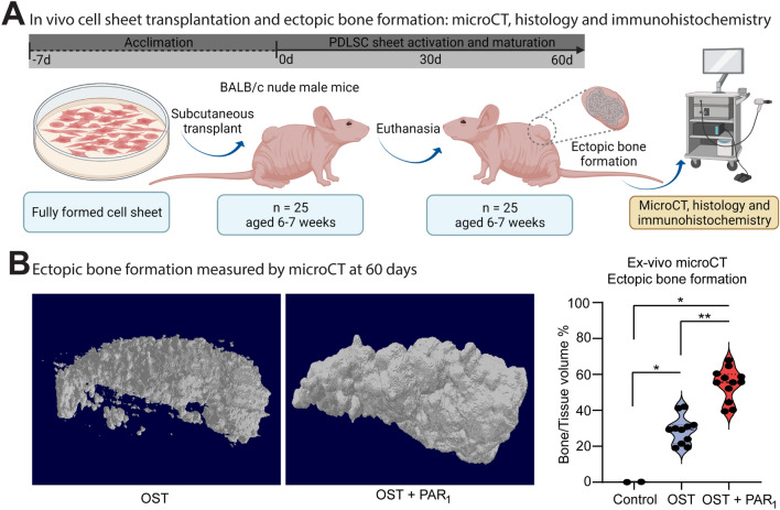
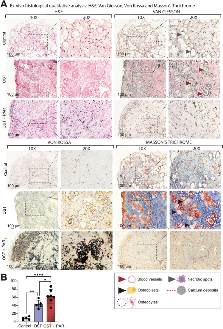
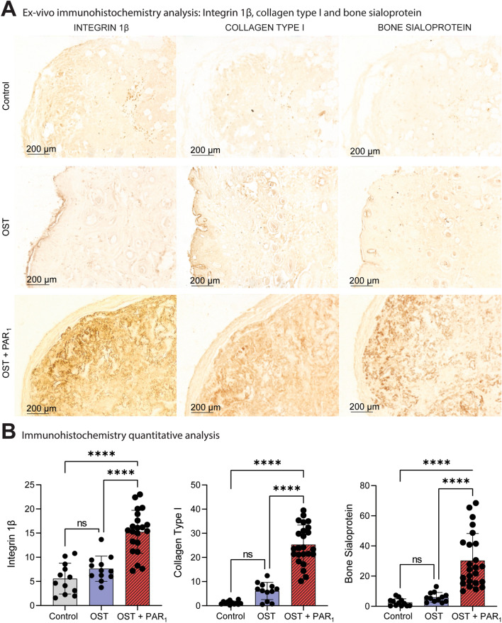
Human periodontal ligament stem cells (PDLSCs) have been studied as a promising strategy in regenerative approaches. The protease-activated receptor 1 (PAR1) plays a key role in osteogenesis and has been shown to induce osteogenesis and increase bone formation in PDLSCs. However, little is known about its effects when activated in PDLSCs as a cell sheet construct and how it would impact bone formation as a graft in vivo. Here, PDLSCs were obtained from 3 patients. Groups were divided into control, osteogenic medium and osteogenic medium + PAR1 activation by TFLLR-NH2 peptide. Cell phenotype was determined by flow cytometry and immunofluorescence. Calcium deposition was quantified by Alizarin Red Staining. Cell sheet microstructure was analyzed through light, scanning electron microscopy and histology and transplanted to Balb/c nude mice. Immunohistochemistry for bone sialoprotein (BSP), integrin β1 and collagen type 1 and histological stains (H&E, Van Giesson, Masson's Trichrome and Von Kossa) were performed on the ex-vivo mineralized tissue after 60 days of implantation in vivo. Ectopic bone formation was evaluated through micro-CT. PAR1 activation increased calcium deposition in vitro as well as BSP, collagen type 1 and integrin β1 protein expression and higher ectopic bone formation (micro-CT) in vivo.


Conflict of interest statement

The authors declare no competing interests.

Figures

Figure 1
Figure 1
(A) Immunofluorescence, flow cytometry and alizarin red staining experiment flow. (B) Flow cytometry analysis showing the phenotypic characterization of PDLSCs for surface stemness, multipotent embryonic biomarkers (SOX2, OCT4, STRO-1) and PAR1 from three individuals (I1, I2 and I3). Unstained cells were used to set positive cell populations, p < 0.05. (C) PAR1 immunofluorescence expression in PDLSCs isolated cell lines. Images in 60X magnification. (D) Alizarin red staining for PDLSCs sheets (I2) after 14 days. Results are given as the mean ± SD. Figure (A) was created using BioRender.
Figure 2
Figure 2
(A) Cell sheets manufacturing and macroscopic morphological characterization workflow. (B) Light microscopy (40x), cell sheet histological profile by H&E staining and microstructural analysis through scanning electron microscopy (400x) from cell sheets for the 3 experimental groups.
Figure 3
Figure 3
(A) Cell sheets in vivo transplant and ex-vivo analysis workflow. (B) Micro-CT analysis of ectopic bone formation in vivo after 60 days of transplantation (B) and the statistical analysis comparing the BV/TV% between the 3 groups CTRL, OST and OST + PAR1. (*) means a p < 0.05 compared to control. Results are given as the mean ± SD. Figure (A) was created using BioRender.
Figure 4
Figure 4
(A) Ex-vivo histological qualitative analysis from H&E, Van Gieson, Masson’s Trichrome and Von Kossa stains from the 3 groups after a 2-month transplantation period in balb/c nude mice. Images at 10x and 20x. CTRL group displayed connective tissue characteristics with necrotic spots (gray arrows) and no bone formation was detected in this group for the 4 stains. Osteoblast clusters (black solid arrows) and blood vessels (red solid arrows) were detected in higher abundance in the OST group when compared with the other groups. The OST + PAR1 group showed greater osteocyte numbers (black dotted circle) and higher calcium deposits (B) (red dotted line).
Figure 5
Figure 5
(A) Histological immunohistochemistry quantification analysis for collagen type I, integrin 1β and bone sialoprotein for the 3 groups ex-vivo samples after a 2-month transplantation period in balb/c nude mice. (B) A semi-quantification analysis was performed for the 3 protein markers and the PAR1 activation group showed greater staining for the 3 osteogenic protein markers after 60 days. Images at 10x. (*) p < 0.05. Results are given as the mean ± SD.

References

    1. Caton G, J., et al. A new classification scheme for periodontal and peri-implant diseases and conditions—Introduction and key changes from the 1999 classification. J. Clin. Periodontol. 2018;45:S1–S8. doi: 10.1111/jcpe.12935. - DOI - PubMed
    1. Magnusson I, Runstad L, Nyman S, Lindhe J. A long junctional epithelium–A locus minoris resistentiae in plaque infection? J. Clin. Periodontol. 1983;10:333–340. doi: 10.1111/j.1600-051X.1983.tb01282.x. - DOI - PubMed
    1. Gao LN, et al. The effect of the coumarin-like derivative osthole on the osteogenic properties of human periodontal ligament and jaw bone marrow mesenchymal stem cell sheets. Biomaterials. 2013;34:9937–9951. doi: 10.1016/j.biomaterials.2013.09.017. - DOI - PubMed
    1. Kim K, Utoh R, Ohashi K, Kikuchi T, Okano T. Fabrication of functional 3D hepatic tissues with polarized hepatocytes by stacking endothelial cell sheets in vitro. J. Tissue Eng. Regen. Med. 2017;11:2071–2080. doi: 10.1002/term.2102. - DOI - PubMed
    1. Mackie EJ, et al. Protease-activated receptors in the musculoskeletal system. Int. J. Biochem. Cell Biol. 2008;40:1169–1184. doi: 10.1016/j.biocel.2007.12.003. - DOI - PubMed

Publication types