. 2023 Jan;95(1):e28157.
doi: 10.1002/jmv.28157. Epub 2022 Sep 28.

Anti-SARS-CoV-2 activity of targeted kinase inhibitors: Repurposing clinically available drugs for COVID-19 therapy

Affiliations

Anti-SARS-CoV-2 activity of targeted kinase inhibitors: Repurposing clinically available drugs for COVID-19 therapy

RuthMabel Boytz et al. J Med Virol. 2023 Jan.

Abstract

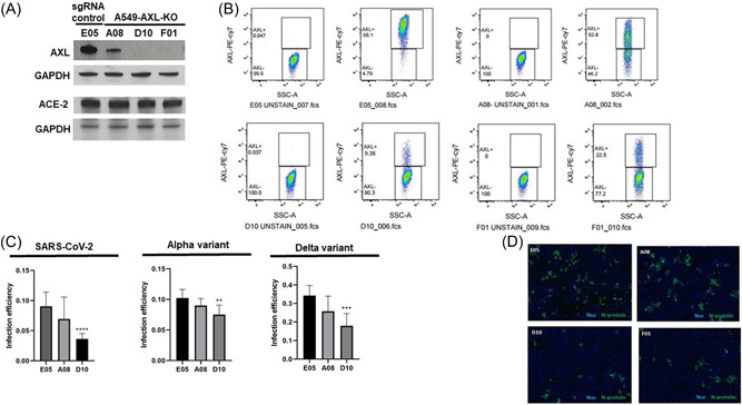
Coronavirus disease 2019 (COVID-19) remains a major public health concern, and vaccine unavailability, hesitancy, or failure underscore the need for discovery of efficacious antiviral drug therapies. Numerous approved drugs target protein kinases associated with viral life cycle and symptoms of infection. Repurposing of kinase inhibitors is appealing as they have been vetted for safety and are more accessible for COVID-19 treatment. However, an understanding of drug mechanism is needed to improve our understanding of the factors involved in pathogenesis. We tested the in vitro activity of three kinase inhibitors against severe acute respiratory syndrome coronavirus 2 (SARS-CoV-2), including inhibitors of AXL kinase, a host cell factor that contributes to successful SARS-CoV-2 infection. Using multiple cell-based assays and approaches, gilteritinib, nintedanib, and imatinib were thoroughly evaluated for activity against SARS-CoV-2 variants. Each drug exhibited antiviral activity, but with stark differences in potency, suggesting differences in host dependency for kinase targets. Importantly, for gilteritinib, the amount of compound needed to achieve 90% infection inhibition, at least in part involving blockade of spike protein-mediated viral entry and at concentrations not inducing phospholipidosis (PLD), approached a clinically achievable concentration. Knockout of AXL, a target of gilteritinib and nintedanib, impaired SARS-CoV-2 variant infectivity, supporting a role for AXL in SARS-CoV-2 infection and supporting further investigation of drug-mediated AXL inhibition as a COVID-19 treatment. This study supports further evaluation of AXL-targeting kinase inhibitors as potential antiviral agents and treatments for COVID-19. Additional mechanistic studies are needed to determine underlying differences in virus response.

Keywords: COVID-19; SARS-CoV-2; antiviral therapy; gilteritinib; imatinib; kinase inhibitor; nintedanib.


Conflict of interest statement

The authors declare no conflict of interest.

Figures

Figure 1
Figure 1
Gilteritinib, imatinib, and nintedanib inhibit SARS‐CoV‐2 infection. (A–C) Representative dose–response curves showing effects of gilteritinib (A), nintedanib (B), and imatinib (C) on SARS‐CoV‐2 infection of A549‐ACE2 cells. One hour before infection cells were treated with a dilution series of each drug starting at 25 µM. Cells were infected with SARS‐CoV‐2 (USA‐WA1 strain) at a multiplicity of infection (MOI) ∼0.2 for 48 h. Cells were fixed and probed for SARS‐CoV‐2 infection using anti‐N protein antibody. Plates were imaged at 4x and infection was quantified using a Cell Profiler pipeline to count the number of N‐positive cells. Infection efficiency, defined as N‐positive cells divided by nuclei, was normalized to a DMSO negative control and dose–response curves and EC50 values were calculated using a four‐parameter variable‐slope model (GraphPad Prism). (D–E) Dose–response curves showing the toxicity of gilteritinib (D) and imatinib (E) on Vero 76 cells. An MOI of 0.001 was used for studies of SARS‐CoV‐2 in Vero 76 cells. The Neutral Red (cytopathic effect/toxicity) assay was carried out as a readout. DMSO, dimethylsulfoxide; ROI, reduction of infection; SARS‐CoV‐2, severe acute respiratory syndrome coronavirus 2.
Figure 2
Figure 2
Kinase inhibitors are less potent against the Beta and Delta strains compared to the Alpha strain. Representative dose–response curves of A549‐ACE2 cells treated with each compound and infected with SARS‐CoV‐2 Alpha, Beta, or Delta strains (A–C). Cells were treated with compounds 1 h before infection with each variant at an MOI ∼0.2. After 48 h of infection plates were fixed and probed for N‐protein. Infection efficiency was calculated by normalizing the proportion of N‐positive cells in each well to an untreated DMSO control. Concentrations listed for each variant are EC50s calculated using a four‐parameter variable slope model (GraphPad Prism). DMSO, dimethylsulfoxide; MOI, multiplicity of infection; SARS‐CoV‐2, severe acute respiratory syndrome coronavirus 2.
Figure 3
Figure 3
SARS‐CoV‐2 infection is reduced in AXL KO cells and correlates with AXL expression level. (A) AXL and ACE‐2 expression assessed by immunoblotting. (B) AXL expression assessed by flow cytometry. (C) A549‐ACE2 cells with AXL knocked out reduced infection efficiency of SARS‐CoV‐2 (left panel), the Alpha variant (middle panel), and the Delta variant (right panel), as compared to control cells (E05). The level of AXL expression correlated with infection inhibition (A08 53%, D10 9%). Graphed results for the Alpha and Delta variant are representative of three independent replicates, for which similar results were observed. AXL expression for the variant studies as measured by flow cytometry is shown in Supporting Information: Figure 1 (D) Representative images A549‐ACE2‐Cas9 cells with AXL knocked down stained with anti‐N protein antibody. Shown are cells infected with SARS‐CoV‐2 at an MOI ∼0.2 for 24 h. Green is N protein, blue is nuclei. Scale bar indicates 300 µM. A549‐ACE2‐Cas9 cells with AXL knocked down were infected with an MOI ∼0.2 for 24 h, fixed in formalin, and probed for infection using an anti‐N protein antibody. Plates were imaged and infection was quantified using a Cell Profiler pipeline to count the number of N‐positive cells. Infection efficiency, defined as N‐positive cells divided by nuclei, was normalized to a DMSO negative control and is shown for the indicated virus variants. Sidak's multiple comparison tests were used to test for significant differences in infection efficiency. Asterisks denote statistical differences versus E05, an AXL‐expressing control line. Data are representative of three or four biological replicates. DMSO, dimethylsulfoxide; MOI, multiplicity of infection; SARS‐CoV‐2, severe acute respiratory syndrome coronavirus 2.
Figure 4
Figure 4
Activity of kinase inhibitors against a panel of pseudotyped viruses. (A) Imatinib, nintedanib, and gilteritinib were tested for antiviral activity in vitro against pseudotyped lentiviruses expressing the spike protein from the SARS‐CoV‐2 (USA‐WA1), Delta, or Omicron strains. Vesicular stomatitis virus (VSV) G protein served as a specificity control. A549‐ACE2 cells were pretreated with the indicated dose of drug and infection was quantified by luciferase expression. Shown are results representative of two biological replicates. (B) The specificity of gilteritinib for SARS‐CoV‐2 and AXL inhibition was tested in vitro using a panel of recombinant VSVs (rVSVs) expressing the Ebola (EBOV), Lassa mammarenavrus (LASV), or VSV glycoproteins. A549‐ACE2 cells were pretreated with gilteritinib and infection was quantified by counting GPF + cells. Concentrations listed are EC50 values calculated using a four‐parameter variable slope model (GraphPad Prism). SARS‐CoV‐2, severe acute respiratory syndrome coronavirus 2.
Figure 5
Figure 5
Effects of gilteritinib, nintedanib, and imatinib on phospholipidosis (PLD) in ACE2‐A549 cells. Comparison of induction of PLD by gilteritinib, nintedanib, and imatinib versus the positive control amodiaquine. Cells were pretreated with NBD‐PE reagent for 2 h before compound addition. Compounds were added in dose curves and cells were incubated overnight. PLD was quantified using Cell Profiler to measure NBD‐PE signal intensity per nuclei, and values were normalized to the negative control DMSO. EC50 values were calculated in Prism using a variable‐slope four‐parameter model.

References

    1. Morse JS, Lalonde T, Xu S, Liu WR. Learning from the past: possible urgent prevention and treatment options for severe acute respiratory infections caused by 2019‐nCoV. ChemBioChem. 2020;21(5):730‐738. 10.1002/cbic.202000047 - DOI - PMC - PubMed
    1. Lu R, Zhao X, Li J, et al. Genomic characterisation and epidemiology of 2019 novel coronavirus: implications for virus origins and receptor binding. Lancet. 2020;395(10224):565‐574. 10.1016/S0140-6736(20)30251-8 - DOI - PMC - PubMed
    1. Shi H, Han X, Jiang N, et al. Radiological findings from 81 patients with COVID‐19 pneumonia in Wuhan, China: a descriptive study. Lancet Infect Dis. 2020;20(4):425‐434. 10.1016/S1473-3099(20)30086-4 - DOI - PMC - PubMed
    1. Gautier JF, Ravussin Y. A new symptom of COVID‐19: loss of taste and smell. Obesity. 2020;28(5):848. 10.1002/oby.22809 - DOI - PMC - PubMed
    1. Jacob JJ, John Fletcher G, Monisha Priya T, Veeraraghavan B, Mutreja A. Relevance of immune response and vaccination strategies of SARS‐CoV‐2 in the phase of viral red queen dynamics. Indian J Med Microbiol. 2021;39(4):417‐422. 10.1016/j.ijmmb.2021.08.001 - DOI - PMC - PubMed

Publication types

Supplementary concepts