

Deficiency of ataxia-telangiectasia mutated kinase attenuates Western-type diet-induced cardiac dysfunction in female mice
- PMID: 36117462
- PMCID: PMC9483716
- DOI: 10.14814/phy2.15434
Deficiency of ataxia-telangiectasia mutated kinase attenuates Western-type diet-induced cardiac dysfunction in female mice
Abstract
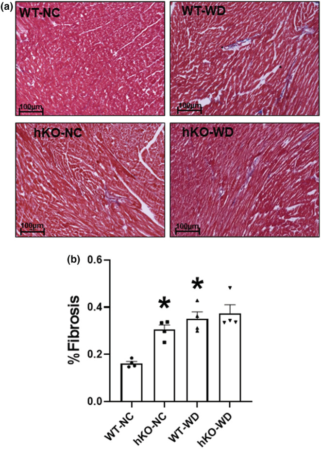
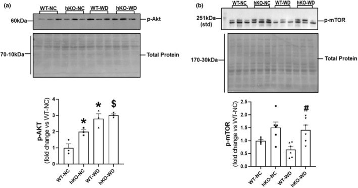
Chronic consumption of Western-type diet (WD) induces cardiac structural and functional abnormalities. Previously, we have shown that WD consumption in male ATM (ataxia-telangiectasia mutated kinase) deficient mice associates with accelerated body weight (BW) gain, cardiac systolic dysfunction with increased preload, and exacerbation of hypertrophy, apoptosis, and inflammation. This study investigated the role of ATM deficiency in WD-induced changes in functional and biochemical parameters of the heart in female mice. Six-week-old wild-type (WT) and ATM heterozygous knockout (hKO) female mice were placed on WD or NC (normal chow) for 14 weeks. BW gain, fat accumulation, and cardiac functional and biochemical parameters were measured 14 weeks post-WD. WD-induced subcutaneous and total fat contents normalized to body weight were higher in WT-WD versus hKO-WD. Heart function measured using echocardiography revealed decreased percent fractional shortening and ejection fraction, and increased LV end systolic diameter and volume in WT-WD versus WT-NC. These functional parameters remained unchanged in hKO-WD versus hKO-NC. Myocardial fibrosis, myocyte hypertrophy, and apoptosis were higher in WT-WD versus WT-NC. However, apoptosis was significantly lower and hypertrophy was significantly higher in hKO-WD versus WT-WD. MMP-9 and Bax expression, and Akt activation were higher in WT-WD versus WT-NC. PARP-1 (full-length) expression and mTOR activation were lower in WT-WD versus hKO-WD. Thus, ATM deficiency in female mice attenuates fat weight gain, preserves heart function, and associates with decreased cardiac cell apoptosis in response to WD.
Keywords: ATM; Western-type diet; apoptosis; female; heart.
© 2022 The Authors. Physiological Reports published by Wiley Periodicals LLC on behalf of The Physiological Society and the American Physiological Society. This article has been contributed to by U.S. Government employees and their work is in the public domain in the USA.
Conflict of interest statement
No conflicts of interest, financial or otherwise, are declared by the authors.
Figures


Similar articles
-
Deficiency of ataxia-telangiectasia mutated kinase modulates functional and biochemical parameters of the heart in response to Western-type diet.Am J Physiol Heart Circ Physiol. 2021 Jun 1;320(6):H2324-H2338. doi: 10.1152/ajpheart.00990.2020. Epub 2021 Apr 30. Am J Physiol Heart Circ Physiol. 2021. PMID: 33929897 Free PMC article.
-
Ataxia telangiectasia-mutated kinase deficiency exacerbates left ventricular dysfunction and remodeling late after myocardial infarction.Am J Physiol Heart Circ Physiol. 2016 Aug 1;311(2):H445-52. doi: 10.1152/ajpheart.00338.2016. Epub 2016 Jun 10. Am J Physiol Heart Circ Physiol. 2016. PMID: 27288435 Free PMC article.
-
Deficiency of ataxia telangiectasia mutated kinase delays inflammatory response in the heart following myocardial infarction.J Am Heart Assoc. 2014 Dec;3(6):e001286. doi: 10.1161/JAHA.114.001286. J Am Heart Assoc. 2014. PMID: 25520329 Free PMC article.
-
Ataxia telangiectasia mutated kinase plays a protective role in β-adrenergic receptor-stimulated cardiac myocyte apoptosis and myocardial remodeling.Mol Cell Biochem. 2011 Jul;353(1-2):13-22. doi: 10.1007/s11010-011-0769-6. Epub 2011 Mar 15. Mol Cell Biochem. 2011. PMID: 21404020 Free PMC article.
-
Poly(ADP-ribose) Polymerase (PARP) and PARP Inhibitors: Mechanisms of Action and Role in Cardiovascular Disorders.Cardiovasc Toxicol. 2018 Dec;18(6):493-506. doi: 10.1007/s12012-018-9462-2. Cardiovasc Toxicol. 2018. PMID: 29968072 Review.
Cited by
-
ATM deficiency differentially affects expression of proteins related to fatty acid oxidation and oxidative stress in a sex-specific manner in response to Western-type diet prior to and following myocardial infarction.Life Sci. 2024 Apr 1;342:122541. doi: 10.1016/j.lfs.2024.122541. Epub 2024 Feb 28. Life Sci. 2024. PMID: 38428572 Free PMC article.
References
-
- Albakri, A. (2018a). Obesity cardiomyopathy: A review of literature on clinical status and meta‐analysis of diagnostic and clinical management. Medical and Clinical Archives, 2, 1–13. 10.15761/MCA.1000134 - DOI
-
- Albakri, A. (2018b). Ischemic heart failure: A review of clinical status and meta‐analysis of diagnosis and clinical management methods. Clinical and Medical Investigations, 3, 1–15. 10.15761/CMI.1000171 - DOI
Publication types
MeSH terms
Substances
Grants and funding
LinkOut - more resources
Full Text Sources
Medical
Research Materials
Miscellaneous
