Case Reports
. 2022 Aug 27:37:89-93.
doi: 10.1016/j.ctro.2022.07.005. eCollection 2022 Nov.

Refractory ventricular tachycardia treated by a second session of stereotactic arrhythmia radioablation

Affiliations
Case Reports

Refractory ventricular tachycardia treated by a second session of stereotactic arrhythmia radioablation

C Herrera Siklody et al. Clin Transl Radiat Oncol. .

Abstract

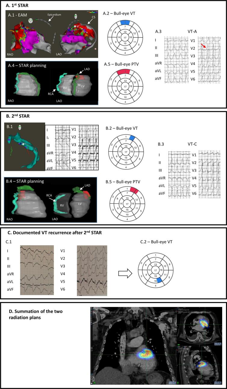
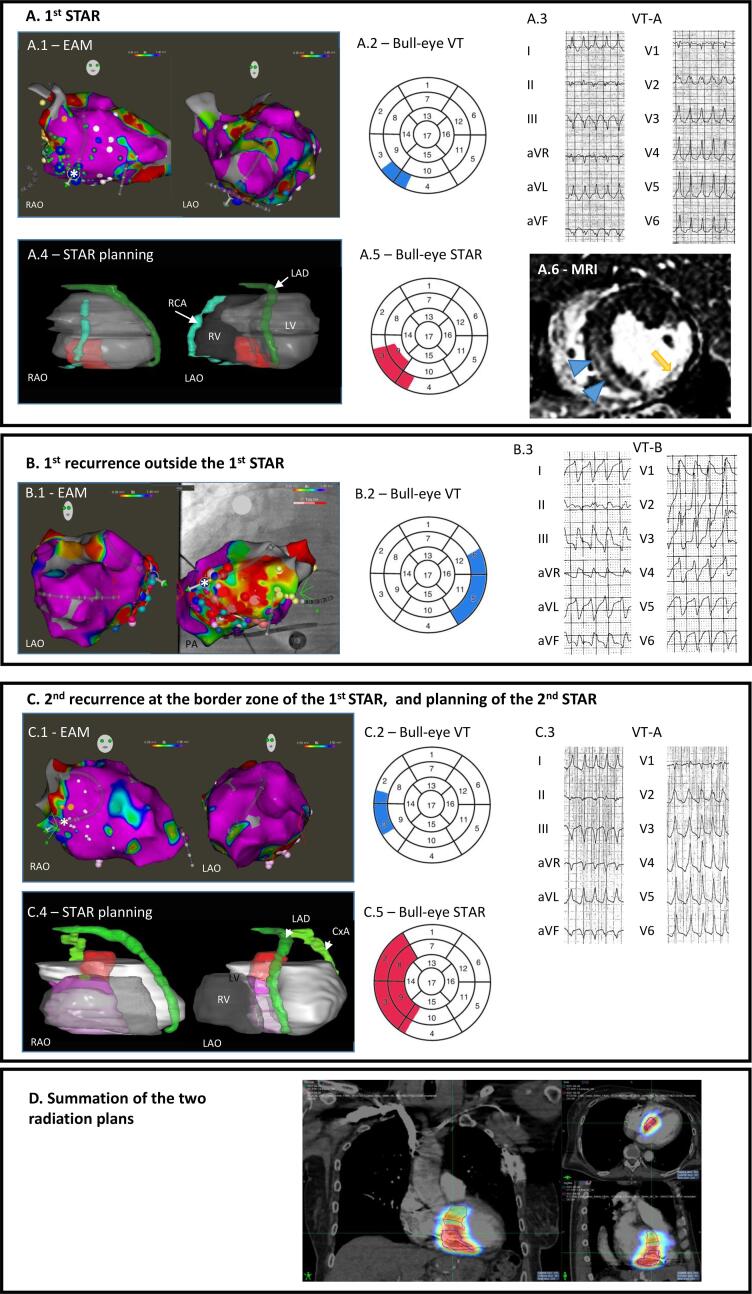
Purpose: Stereotactic arrhythmia radioablation (STAR) is an effective treatment for refractory ventricular tachycardia (VT), but recurrences after STAR were recently published. Herein, we report two cases of successful re-irradiation of the arrhythmogenic substrate.

Cases: We present two cases of re-irradiation after recurrence of a previously treated VT with radioablation at a dose of 20 Gy. The VT exit was localized on the border zone of the irradiated volume, which responded positively to re-irradiation at follow-up.

Conclusion: These two cases show the technical feasibility of re-irradiation to control recurrent VT after a first STAR.

Keywords: Ischemic heart disease; Non-ischemic cardiomyopathy; Stereotactic arrhythmia radioablation (STAR); ventricular tachycardia (VT).


Conflict of interest statement

The authors declare that they have no known competing financial interests or personal relationships that could have appeared to influence the work reported in this paper.

Figures

Fig. 1
Fig. 1
Overview of the procedures for Case 1. A.1. Epicardial anterior VT targeted by STAR. EAM of the LV during the last CA before STAR: bipolar voltage maps in RAO (left) and RAO (right) views, with the tip of the ablation catheter (white star) located in the CS at the best pacemap spot. A decapolar diagnostic catheter is positioned epicardially facing the ablation catheter. A.2. Bull eye’s plot displaying the VT substrate location in the 17-segment AHA model (in blue). A.3. 12-lead ECG of VT-A, displaying a pseudo-delta wave (red arrow) suggesting an epicardial exit. A.4. RAO and LAO views of the STAR plan with the PTV in red. A.5. Bull eye’s plot displaying the location of the PTV in the 17-segment AHA model (in red). B.1. Second STAR procedure. EAM of the coronary sinus corresponding to the last CA before the 2nd STAR with the tip of the ablation catheter (white star) located at the best pacemap spot. Note that the ablation catheter lies more laterally as compared to its 1st location in figure 1A. Unipolar pacemapping with a guidewire is displayed on the right. B.2. Bull eye’s plot displaying the location of the VT exit in the 17-segment AHA-model (in blue). B.3. 12-lead ECG of the induced VT (VT-C). B.4. RAO and LAO views of the STAR plan with the PTV in red. B.5. Bull eye’s plot displaying the location of the PTV in the 17-segment AHA-model (in red). C. VT from the ischemic scar observed in 2022. C.1. 12-lead ECG and C.2. location on the 17-segment AHA-model (in blue) of the exit of the documented VT recurrence after the 2nd STAR procedure. RAO: right anterior oblique; LAO: left anterior oblique; CS: coronary sinus; RCA: right coronary artery; LAD: left anterior descending coronary artery: RV: right ventricle; LV: left ventricle. D. Summation of the two radiation plans.
Fig. 2
Fig. 2
Overview of the procedures for Case 2. A.1. VT from the septal fibrotic infiltration targeted by STAR. EAM of the LV from the last CA before STAR: bipolar voltage maps in RAO (left) and RAO (right) views, with the tip of the ablation catheter (white star) located at the best endocardial pacemap spot corresponding to the inferior basal IVS (blue tags). A.2. Bull eye’s view displaying the exit of the VT in the 17-segment AHA-model (in blue). A.3. 12-lead ECG of the clinical VT (VT-A). A.4. 3-D reconstruction of the irradiation plan with the PTV in red. A.5. Bull eye’s plot displaying the location of the PTV based on the 17-segment AHA-model (in red). A.6. Late enhancement cardiac MRI showing the fibrotic infiltration of the septum (blue arrowheads) and of the infero-lateral MI (yellow arrow). B.1. VT from the ischemic scar successfully treated by CA. LV EAM following the 1st VT recurrence after STAR: bipolar voltage maps in LAO (left) and PA (right) views, with the tip of the ablation catheter (white star) located at the successful ablation site. Red tags indicate ablation spots. B.2. Bull eye’s plot displaying the location of the VT substrate in the 17-segment AHA-model (in blue). B.3. 12-lead ECG of the induced VT (VT-B) that differed from VT-A shown in panel A3. C.1. Second STAR procedure targeting the septal non-ischemic VT. LV EAM corresponding to the last CA before the 2nd STAR procedure: bipolar voltage maps in RAO (left) and RAO (right) views, with the tip of the ablation catheter (white star) located at the best endocardial pacemap spot at the intersection of the inferior and middle third of the basal IVS. C.2. Bull eye’s plot displaying the location of VT exit in the 17-segment AHA-model (in blue). C.3. 12-lead ECG of the clinical VT (VT-A). C.4. RAO and LAO views of the STAR plan with the PTV in red and the complementary and transition volume in pink and fuchsia respectively. C.5. Bull eye’s plot displaying the location of the new PTV in the 17-segment AHA-model (in red). RAO: right anterior oblique; LAO: left anterior oblique; PA: postero-anterior; CS: coronary sinus; RCA: right coronary artery; LAD: left anterior descending coronary artery: CxA: circumflex coronary artery; RV: right ventricle; LV: left ventricle. D. Summation of the two radiation plans.

References

    1. Cuculich P.S., Schill M.R., Kashani R., Mutic S., Lang A., Cooper D., et al. Noninvasive cardiac radiation for ablation of ventricular tachycardia. N Engl J Med. 2017;377(24):2325–2336. - PMC - PubMed
    1. Neuwirth R., Cvek J., Knybel L., Jiravsky O., Molenda L., Kodaj M., et al. Stereotactic radiosurgery for ablation of ventricular tachycardia. Europace. 2019;21:1088–1095. - PubMed
    1. Lloyd M.S., Wight J., Schneider F., Hoskins M., Attia T., Escott C., et al. Clinical experience of stereotactic body radiation for refractory ventricular tachycardia in advanced heart failure patients. Heart Rhythm. 2020;17(3):415–422. - PubMed
    1. Gianni C., Rivera D., Burkhardt J.D., Pollard B., Gardner E., Maguire P., et al. Stereotactic arrhythmia radioablation for refractory scar-related ventricular tachycardia. Heart Rhythm. 2020;17(8):1241–1248. - PubMed
    1. Jumeau R., Ozsahin M., Schwitter J., Vallet V., Duclos F., Zeverino M., et al. Rescue procedure for an electrical storm using robotic non-invasive cardiac radio-ablation. Radiother Oncol. 2018;128(2):189–191. - PubMed

Publication types

LinkOut - more resources