Downregulation of AKT/mTOR signaling pathway for Salmonella-mediated autophagy in human anaplastic thyroid cancer
- PMID: 36118522
- PMCID: PMC9475365
- DOI: 10.7150/jca.75163
Downregulation of AKT/mTOR signaling pathway for Salmonella-mediated autophagy in human anaplastic thyroid cancer
Abstract
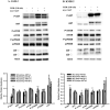
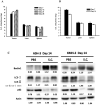
Thyroid cancer has been known as the most common endocrine malignancy. Although majority of thyroid cancer types respond well to conventional treatment including surgery and radioactive iodine therapy, about 10% of those with differentiated thyroid cancer will present distant metastasis and will have persistent or recurrent disease. Even more serious is a rare type of thyroid cancer called anaplastic thyroid cancer (ATC), which accounts for about 1%, has been demonstrated as the most lethal and aggressive form of human malignancy. Unfortunately, these tumors are also frequently resistant to traditional therapy. Previous study have shown that Salmonella inhibits tumor growth, in part, by inducing autophagy - a cellular process that is important in the innate and adaptive immunity in response to viral or bacterial infection. In our study, we intended to investigate whether Salmonella can inhibit tumor growth by inducing autophagy, specifically in thyroid cancer and elucidate the possible molecular mechanism. In order to determine the signaling pathway involved in tumor cell autophagy, we used Salmonella to treat ATC cells line ASH-3 and KMH-2 in vitro. The autophagic markers, particularly autophagy-related gene 6 (Beclin-1), microtubule-associated protein 1A/1B-light chain 3 (LC3) and p62, were observed to be differentially expressed after infection with Salmonella indicating an activated autophagy in ATC cells. In addition, the protein expression levels of phospho-protein kinase B (P-AKT), phospho-mammalian targets of rapamycin (P-mTOR), phospho-p70 ribosomal s6 kinase (P-p70S6K) in tumor cells were decreased after Salmonella infection. In vivo, we also found that substantial cell numbers of Salmonella targeted tumor tissue, and regulated anti-tumor mechanisms. Our findings showed that Salmonella activated autophagic signaling pathway and inhibited ATC tumor growth via downregulation of AKT/mTOR pathway.
Keywords: AKT/mTOR; Autophagy; Salmonella; anaplastic thyroid cancer.
© The author(s).
Conflict of interest statement
Competing Interests: The authors have declared that no competing interest exists.
Figures


Similar articles
-
Anaplastic thyroid cancer: Genetic roles, targeted therapy, and immunotherapy.Genes Dis. 2024 Aug 30;12(4):101403. doi: 10.1016/j.gendis.2024.101403. eCollection 2025 Jul. Genes Dis. 2024. PMID: 40271195 Free PMC article. Review.
-
Salmonella induce autophagy in melanoma by the downregulation of AKT/mTOR pathway.Gene Ther. 2014 Mar;21(3):309-16. doi: 10.1038/gt.2013.86. Epub 2014 Jan 23. Gene Ther. 2014. PMID: 24451116
-
Digitalislike Compounds Restore hNIS Expression and Iodide Uptake Capacity in Anaplastic Thyroid Cancer.J Nucl Med. 2018 May;59(5):780-786. doi: 10.2967/jnumed.117.200675. Epub 2017 Dec 14. J Nucl Med. 2018. PMID: 29242405
-
Simultaneous activation of impaired autophagy and the mammalian target of rapamycin pathway in oral squamous cell carcinoma.J Oral Pathol Med. 2019 Sep;48(8):705-711. doi: 10.1111/jop.12884. Epub 2019 Jun 17. J Oral Pathol Med. 2019. PMID: 31132188
-
Long Non-Coding RNAs as Determinants of Thyroid Cancer Phenotypes: Investigating Differential Gene Expression Patterns and Novel Biomarker Discovery.Biology (Basel). 2024 Apr 27;13(5):304. doi: 10.3390/biology13050304. Biology (Basel). 2024. PMID: 38785786 Free PMC article. Review.
Cited by
-
Transcriptomic and proteomic analysis of the jejunum revealed the effects and mechanism of protocatechuic acid on alleviating Salmonella typhimurium infection in chickens.Poult Sci. 2025 Jan;104(1):104606. doi: 10.1016/j.psj.2024.104606. Epub 2024 Nov 27. Poult Sci. 2025. PMID: 39631287 Free PMC article.
-
Anaplastic thyroid cancer: Genetic roles, targeted therapy, and immunotherapy.Genes Dis. 2024 Aug 30;12(4):101403. doi: 10.1016/j.gendis.2024.101403. eCollection 2025 Jul. Genes Dis. 2024. PMID: 40271195 Free PMC article. Review.
-
Salmonella enterica and outer membrane vesicles are current and future options for cancer treatment.Front Cell Infect Microbiol. 2023 Dec 5;13:1293351. doi: 10.3389/fcimb.2023.1293351. eCollection 2023. Front Cell Infect Microbiol. 2023. PMID: 38116133 Free PMC article. Review.
-
Gut Microbiota-Tumor Microenvironment Interactions: Mechanisms and Clinical Implications for Immune Checkpoint Inhibitor Efficacy in Cancer.Cancer Manag Res. 2025 Jan 25;17:171-192. doi: 10.2147/CMAR.S405590. eCollection 2025. Cancer Manag Res. 2025. PMID: 39881948 Free PMC article. Review.
-
Arbutin overcomes tumor immune tolerance by inhibiting tumor programmed cell death-ligand 1 expression.Int J Med Sci. 2024 Nov 11;21(15):2992-3002. doi: 10.7150/ijms.92419. eCollection 2024. Int J Med Sci. 2024. PMID: 39628695 Free PMC article.
References
-
- Mirian C, Gronhoj C, Jensen DH, Jakobsen KK, Karnov K, Jensen JS. et al. Trends in thyroid cancer: Retrospective analysis of incidence and survival in Denmark 1980-2014. Cancer Epidemiol. 2018;55:81–7. - PubMed
-
- Mihailovic J, Stefanovic L, Malesevic M. Differentiated Thyroid Carcinoma With Distant Metastases: Probability of Survival and Its Predicting Factors. Cancer Biother Radiopharm. 2007;22:250–5. - PubMed
LinkOut - more resources
Full Text Sources
Research Materials
Miscellaneous

