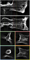
Location of pedicle screw hold in relation to bone quality and loads
- PMID: 36118575
- PMCID: PMC9478651
- DOI: 10.3389/fbioe.2022.953119
Location of pedicle screw hold in relation to bone quality and loads
Abstract
Introduction: Sufficient screw hold is an indispensable requirement for successful spinal fusion, but pedicle screw loosening is a highly prevalent burden. The aim of this study was to quantify the contribution of the pedicle and corpus region in relation to bone quality and loading amplitude of pedicle screws with traditional trajectories. Methods: After CT examination to classify bone quality, 14 pedicle screws were inserted into seven L5. Subsequently, Micro-CT images were acquired to analyze the screw's location and the vertebrae were split in the midsagittal plane and horizontally along the screw's axis to allow imprint tests with 6 mm long sections of the pedicle screws in a caudal direction perpendicular to the screw's surface. Force-displacement curves in combination with the micro-CT data were used to reconstruct the resistance of the pedicle and corpus region at different loading amplitudes. Results: Bone quality was classified as normal in three specimens, as moderate in two and as bad in two specimens, resulting in six, four, and four pedicle screws per group. The screw length in the pedicle region in relation to the inserted screw length was measured at an average of 63%, 62%, and 52% for the three groups, respectively. At a calculated 100 N axial load acting on the whole pedicle screw, the pedicle region contributed an average of 55%, 58%, and 58% resistance for the normal, moderate, and bad bone quality specimens, respectively. With 500 N load, these values were measured at 59%, 63%, and 73% and with 1000 N load, they were quantified at 71%, 75%, and 81%. Conclusion: At lower loading amplitudes, the contribution of the pedicle and corpus region on pedicle screw hold are largely balanced and independent of bone quality. With increasing loading amplitudes, the contribution of the pedicle region increases disproportionally, and this increase is even more pronounced in situations with reduced bone quality. These results demonstrate the importance of the pedicle region for screw hold, especially for reduced bone quality.
Keywords: bone density; instrumentation; lumbar spine; pedicle screw; primary stability; screw bone interface.
Copyright © 2022 Cornaz, Farshad and Widmer.
Conflict of interest statement
The authors declare that the research was conducted in the absence of any commercial or financial relationships that could be construed as a potential conflict of interest.
Figures


Similar articles
-
Bone density optimized pedicle screw instrumentation improves screw pull-out force in lumbar vertebrae.Comput Methods Biomech Biomed Engin. 2022 Mar;25(4):464-474. doi: 10.1080/10255842.2021.1959558. Epub 2021 Aug 9. Comput Methods Biomech Biomed Engin. 2022. PMID: 34369827
-
Effect of augmentation techniques on the failure of pedicle screws under cranio-caudal cyclic loading.Eur Spine J. 2017 Jan;26(1):181-188. doi: 10.1007/s00586-015-3904-3. Epub 2015 Mar 27. Eur Spine J. 2017. PMID: 25813011
-
Biomechanical evaluation of pedicle screw loosening mechanism using synthetic bone surrogate of various densities.Annu Int Conf IEEE Eng Med Biol Soc. 2014;2014:4346-9. doi: 10.1109/EMBC.2014.6944586. Annu Int Conf IEEE Eng Med Biol Soc. 2014. PMID: 25570954
-
Patient-Specific Finite Element Models of Posterior Pedicle Screw Fixation: Effect of Screw's Size and Geometry.Front Bioeng Biotechnol. 2021 Mar 10;9:643154. doi: 10.3389/fbioe.2021.643154. eCollection 2021. Front Bioeng Biotechnol. 2021. PMID: 33777914 Free PMC article.
-
Pedicle screw placement in the lumbar spine: effect of trajectory and screw design on acute biomechanical purchase.J Neurosurg Spine. 2015 May;22(5):503-10. doi: 10.3171/2014.10.SPINE14205. Epub 2015 Feb 13. J Neurosurg Spine. 2015. PMID: 25679236
Cited by
-
A Novel Free-hand Technique of Pedicle Screw Placement in the Lumbar Spine: Accuracy Evaluation and Preliminary Clinical Results.Orthop Surg. 2023 Sep;15(9):2260-2266. doi: 10.1111/os.13750. Epub 2023 Jul 21. Orthop Surg. 2023. PMID: 37476856 Free PMC article.
-
Two-Stage Lumbar Dynamic Stabilization Surgery: A Comprehensive Analysis of Screw Loosening Rates and Functional Outcomes Compared to Single-Stage Approach in Osteopenic and Osteoporotic Patients.Diagnostics (Basel). 2024 Jul 12;14(14):1505. doi: 10.3390/diagnostics14141505. Diagnostics (Basel). 2024. PMID: 39061642 Free PMC article.
-
Visuohaptic Feedback in Robotic-Assisted Spine Surgery for Pedicle Screw Placement.J Clin Med. 2025 May 29;14(11):3804. doi: 10.3390/jcm14113804. J Clin Med. 2025. PMID: 40507566 Free PMC article.
-
Assessment of the tolerance angle for pedicle screw insertion.Med Biol Eng Comput. 2024 Apr;62(4):1265-1275. doi: 10.1007/s11517-023-03002-x. Epub 2024 Jan 4. Med Biol Eng Comput. 2024. PMID: 38177833
-
Hounsfield unit for assessing bone mineral density distribution within lumbar vertebrae and its clinical values.Front Endocrinol (Lausanne). 2024 Jun 13;15:1398367. doi: 10.3389/fendo.2024.1398367. eCollection 2024. Front Endocrinol (Lausanne). 2024. PMID: 38938515 Free PMC article.
References
-
- Afifi M. B., Abdelrazek A., Deiab N. A., Abd El-Hafez A. I., El-Farrash A. H. (2020). The effects of CT x-ray tube voltage and current variations on the relative electron density (RED) and CT number conversion curves. J. Radiat. Res. Appl. Sci. 13 (1), 1–11. 10.1080/16878507.2019.1693176 - DOI
-
- Aichmair A., Moser M., Bauer M. R., Bachmann E., Snedeker J. G., Betz M., et al. (2017). Pull-out strength of patient-specific template-guided vs. free-hand fluoroscopically controlled thoracolumbar pedicle screws: a biomechanical analysis of a randomized cadaveric study. Eur. Spine J. 26 (11), 2865–2872. 10.1007/s00586-017-5025-7 - DOI - PubMed
-
- Bartel D. L., Davy D. T., Keaveny T. M. (2006). Orthopaedic Biomechanics: Mechanics and design in musculoskeletal systems. Upper Saddle River, New Jersey: Prentice-Hall, 370. First edit. Prentice Hall.
LinkOut - more resources
Full Text Sources
Research Materials

