. 2022 Jul-Aug;119(4):343-350.

Thyroid Eye Disease

Affiliations

Thyroid Eye Disease

Jason Szelog et al. Mo Med. 2022 Jul-Aug.

Abstract

Thyroid eye disease, although rare, is the most common inflammatory orbital disorder and is associated with autoimmune thyroid dysfunction. It is a progressive disorder with symptoms and signs that may cause significant facial disfigurement, visual disability, but rarely blindness. We will review the diagnostic criteria, immunologic basis, clinical course, and medical and surgical treatments for thyroid eye disease. Recent developments in the use of biologic agents to treat this disorder appear to be changing its progression curve and offer the first specific and preventative therapeutic options.


Conflict of interest statement

Disclosure MCS is a Consultant/Advisor to Horizon Therapeutics.

Figures

Figures 1A and B
Figures 1A and B
A 45-year-old male with active TED demonstrating severe proptosis, upper and lower eyelid retraction and conjunctival injection and edema.
Figures 2 A and B
Figures 2 A and B
Axial (A) and coronal (B) CT images of a 65-year-old female smoker with compressive optic neuropathy. Note severe enlargement of all rectus muscles impinging on the optic nerves in the retrobulbar orbit.
Figures 3 A and B
Figures 3 A and B
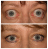
Pre (A) and post (B) teprotumumab images for a 67-year-old female. Her CAS reduced from 4 to 0 and there was a 3 mm reduction in her axial proptosis of the right eye.
Figure 4
Figure 4
Schematic of the orbital bones commonly removed in orbital decompression surgery. This is a right orbit with medial wall outlined in blue, orbital floor in orange and lateral wall in yellow. Reprinted with permission from the American Academy of Ophthalmology 2020–21 BCSC Basic and Clinical Science Book.
Figures 5 A and B
Figures 5 A and B
(A) A 50-year-old female with inactive TED with proptosis and upper and lower eyelid retraction. (B) After bilateral orbital bony balanced decompression of the medial and lateral orbital walls.
None

References

    1. Bartley GB, Gorman CA. Diagnostic criteria for Graves’ ophthalmopathy. Am J Ophthalmol. 1995 Jun;119(6):792–5. - PubMed
    1. Li Z, Cestari DM, Fortin E. Thyroid eye disease: what is new to know? Curr Opin Ophthalmol. 2018 Nov;29(6):528–534. - PubMed
    1. Meyer P, Das T, Ghadiri N, Murthy R, Theodoropoulou S. Clinical pathophysiology of thyroid eye disease: The Cone Model. Eye (Lond) 2019 Feb;33(2):244–253. - PMC - PubMed
    1. Douglas RS, Afifiyan NF, Hwang CJ, et al. Increased generation of fibrocytes in thyroid-associated ophthalmopathy. J Clin Endocrinol Metab. 2010 Jan;95(1):430–8. - PMC - PubMed
    1. Lazarus JH. Epidemiology of Graves’ orbitopathy (GO) and relationship with thyroid disease. Best Pract Res Clin Endocrinol Metab. 2012 Jun;26(3):273–9. - PubMed

Substances

LinkOut - more resources