

CX3CL1 Derived from Bone Marrow Mesenchymal Stem Cells Inhibits A β 1-42-Induced SH-SY5Y Cell Pathological Damage through TXNIP/NLRP3 Signaling Pathway
- PMID: 36118839
- PMCID: PMC9477634
- DOI: 10.1155/2022/1949344
CX3CL1 Derived from Bone Marrow Mesenchymal Stem Cells Inhibits A β 1-42-Induced SH-SY5Y Cell Pathological Damage through TXNIP/NLRP3 Signaling Pathway
Retraction in
-
Retracted: CX3CL1 Derived from Bone Marrow Mesenchymal Stem Cells Inhibits Aβ1-42-Induced SH-SY5Y Cell Pathological Damage through TXNIP/NLRP3 Signaling Pathway.Comput Math Methods Med. 2023 Jun 28;2023:9817614. doi: 10.1155/2023/9817614. eCollection 2023. Comput Math Methods Med. 2023. PMID: 37416293 Free PMC article.
Abstract
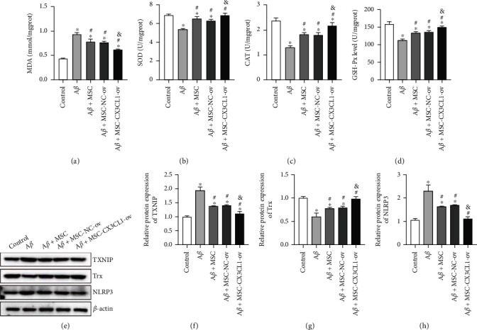
Alzheimer's disease (AD) is the most commonly seen neurodegenerative brain disorder. The paracrine effects of mesenchymal stem cells (MSCs) signify to trigger immunomodulation and neural regeneration. However, the role and mechanism of bone marrow MSC- (BMSC-) derived CX3CL1 in AD remains elusive. In this study, Aβ 1-42-intervened SH-SY5Y cells were used for AD cell model construction. pcDNA-ligated CX3CL1 overexpression plasmids were transfected into BMSCs. The levels of soluble and membrane-bound CX3CL1 were detected by ELISA and Western blotting (WB), respectively. The growth, apoptosis, and pathology of AD model cells were evaluated by CCK-8, flow cytometry, immunofluorescence, morphology observation, biochemical examination, and WB. It was found that Aβ 1-42 significantly reduced CX3CL1 expression either in soluble or membrane-bound form, cell viability, relative protein expression of synaptic markers, SOD, CAT, and GSH-Px contents, as well as Trx protein expression; in addition, it enhanced the apoptosis rate, the relative expression of cleaved caspase-3, Aβ, tau, p-Tau, Iba1, MDA, TXNIP, and NLRP3 in SH-SY5Y cells; however, the above effects were prominently reversed by the coculture of BMSCs. Moreover, overexpression of CX3CL1 in BMSCs observably strengthened the corresponding tendency caused by BMSCs. In conclusion, through the TXNIP/NLRP3 pathway, CX3CL1 derived from BMSCs inhibited pathological damage in Aβ 1-42-induced SH-SY5Y.
Copyright © 2022 Chuan Guo et al.
Conflict of interest statement
The authors report that there are no competing interests to declare.
Figures


Similar articles
-
Dl-3-n-Butylphthalide Inhibits NLRP3 Inflammasome and Mitigates Alzheimer's-Like Pathology via Nrf2-TXNIP-TrX Axis.Antioxid Redox Signal. 2019 Apr 10;30(11):1411-1431. doi: 10.1089/ars.2017.7440. Epub 2018 Apr 25. Antioxid Redox Signal. 2019. PMID: 29634349
-
Inhibitor of RAGE and glucose‑induced inflammation in bone marrow mesenchymal stem cells: Effect and mechanism of action.Mol Med Rep. 2020 Oct;22(4):3255-3262. doi: 10.3892/mmr.2020.11422. Epub 2020 Aug 7. Mol Med Rep. 2020. PMID: 32945430 Free PMC article.
-
Inhibition of endotoxin-induced acute lung injury in rats by bone marrow-derived mesenchymal stem cells: Role of Nrf2/HO-1 signal axis in inhibition of NLRP3 activation.Biochem Biophys Res Commun. 2021 Apr 30;551:7-13. doi: 10.1016/j.bbrc.2021.03.009. Epub 2021 Mar 10. Biochem Biophys Res Commun. 2021. PMID: 33713981
-
Gclc overexpression inhibits apoptosis of bone marrow mesenchymal stem cells through the PI3K/AKT/Foxo1 pathway to alleviate inflammation in acute lung injury.Int Immunopharmacol. 2022 Sep;110:109017. doi: 10.1016/j.intimp.2022.109017. Epub 2022 Jul 2. Int Immunopharmacol. 2022. PMID: 35792274
-
Effects of CX3CR1 and Fractalkine Chemokines in Amyloid Beta Clearance and p-Tau Accumulation in Alzheimer's Disease (AD) Rodent Models: Is Fractalkine a Systemic Biomarker for AD?Curr Alzheimer Res. 2016;13(4):403-12. doi: 10.2174/1567205013666151116125714. Curr Alzheimer Res. 2016. PMID: 26567742 Review.
Cited by
-
CX3CL1 Pathway as a Molecular Target for Treatment Strategies in Alzheimer's Disease.Int J Mol Sci. 2023 May 4;24(9):8230. doi: 10.3390/ijms24098230. Int J Mol Sci. 2023. PMID: 37175935 Free PMC article. Review.
-
Retracted: CX3CL1 Derived from Bone Marrow Mesenchymal Stem Cells Inhibits Aβ1-42-Induced SH-SY5Y Cell Pathological Damage through TXNIP/NLRP3 Signaling Pathway.Comput Math Methods Med. 2023 Jun 28;2023:9817614. doi: 10.1155/2023/9817614. eCollection 2023. Comput Math Methods Med. 2023. PMID: 37416293 Free PMC article.
References
-
- Wiley J. Alzheimer’s disease facts and figures. Alzheimers Dementia . 2021;17 - PubMed
Publication types
MeSH terms
Substances
LinkOut - more resources
Full Text Sources
Medical
Research Materials
Miscellaneous
