. 2022 Sep 2:13:948047.
doi: 10.3389/fimmu.2022.948047. eCollection 2022.

Lyl1-deficiency promotes inflammatory responses and increases mycobacterial burden in response to Mycobacterium tuberculosis infection in mice

Affiliations

Lyl1-deficiency promotes inflammatory responses and increases mycobacterial burden in response to Mycobacterium tuberculosis infection in mice

Shelby-Sara Jones et al. Front Immunol. .

Abstract

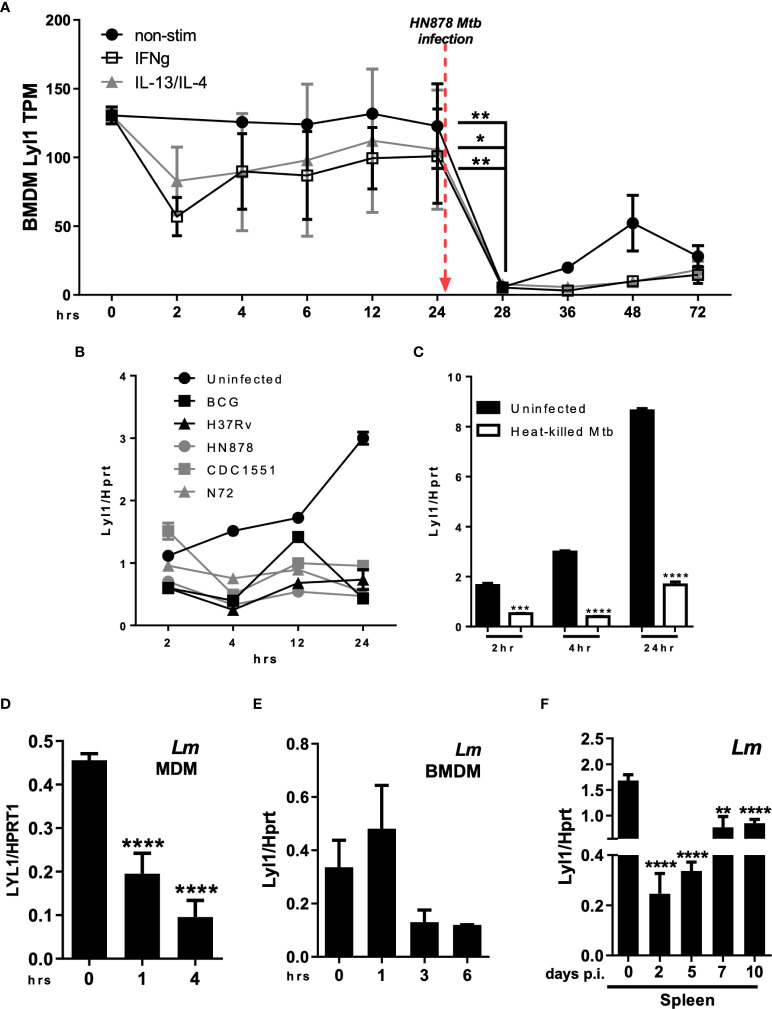
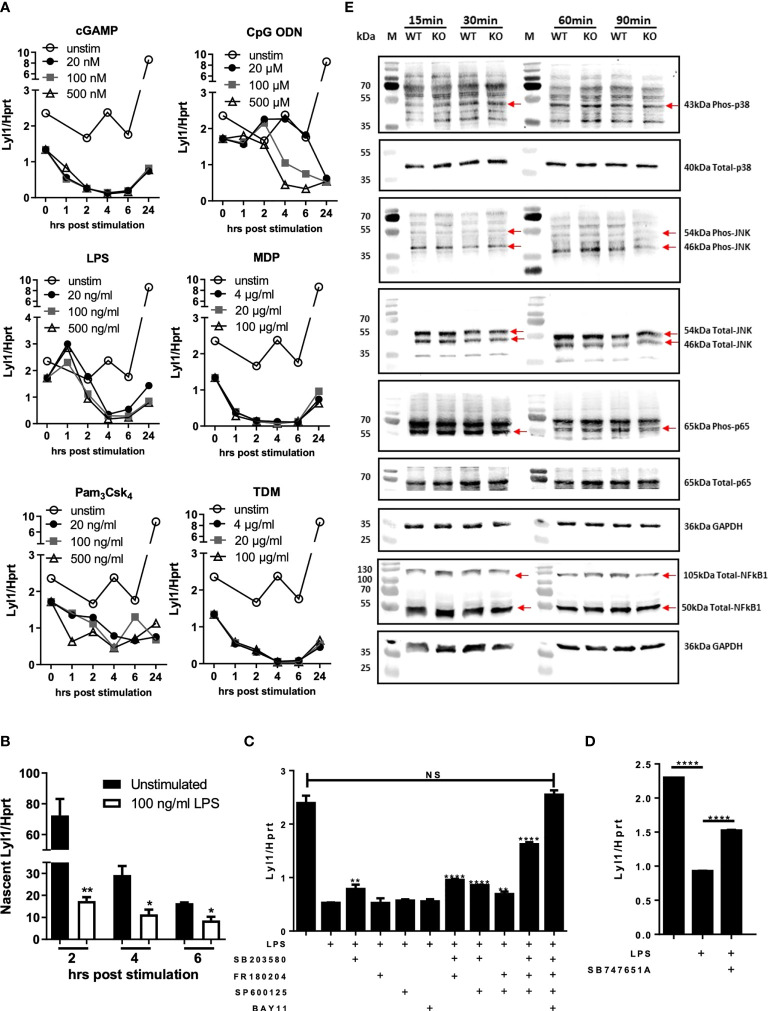
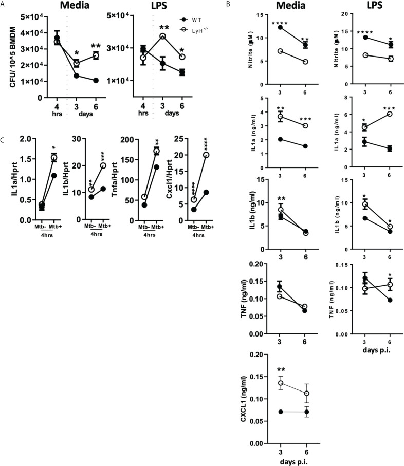
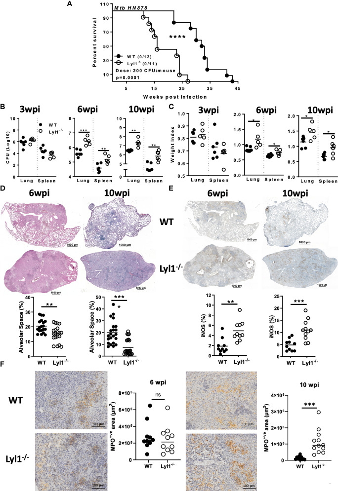
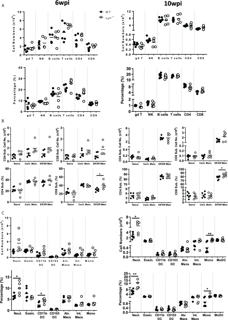
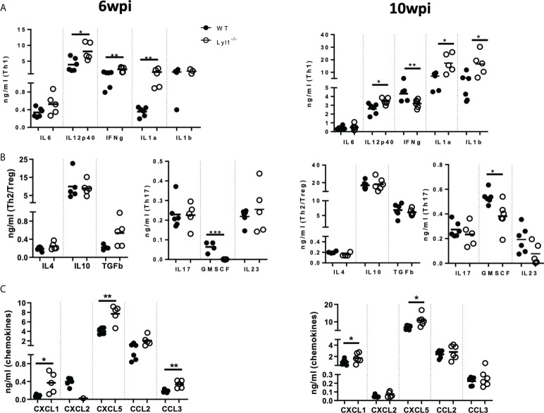
Lymphoblastic leukemia 1 (Lyl1) is a well-studied transcription factor known to exhibit oncogenic potential in various forms of leukemia with pivotal roles in hematopoietic stem cell biology. While its role in early hematopoiesis is well established, its function in mature innate cells is less explored. Here, we identified Lyl1 as a drastically perturbed gene in the Mycobacterium tuberculosis (Mtb) infected mouse macrophage transcriptome. We report that Lyl1 downregulation upon immune stimulation is a host-driven process regulated by NFκB and MAP kinase pathways. Interestingly, Lyl1-deficient macrophages have decreased bacterial killing potential with reduced nitric oxide (NO) levels while expressing increased levels of pro-inflammatory interleukin-1 and CXCL1. Lyl1-deficient mice show reduced survival to Mtb HN878 infection with increased bacterial burden and exacerbated inflammatory responses in chronic stages. We observed that increased susceptibility to infection was accompanied by increased neutrophil recruitment and IL-1, CXCL1, and CXCL5 levels in the lung homogenates. Collectively, these results suggest that Lyl1 controls Mtb growth, reduces neutrophilic inflammation and reveals an underappreciated role for Lyl1 in innate immune responses.

Keywords: Mycobacterium tuberculosis; innate immunity; lymphoblastic leukemia 1; neutrophilic inflammation; transcription factor.


Conflict of interest statement

The authors declare that the research was conducted in the absence of any commercial or financial relationships that could be construed as a potential conflict of interest.

Figures

Figure 1
Figure 1
Regulation of Lyl1 expression is a host-driven mechanism. (A) Expression kinetics (represented as Tags Per Million (TPM)) of Lyl1 in Mtb HN878 infected mouse bone marrow-derived macrophage (BMDM) data were extracted from the FANTOM5 mouse macrophages dataset. (B) Lyl1 mRNA expression was measured by RT-qPCR, relative to the Hprt housekeeping gene, in BMDM infected with various Mtb strains, as well as (C) heat-killed Mtb at MOI:1. (D) MDM derived from healthy participants were infected with Lm with MOI:10 to measure LYL1 mRNA expression kinetics. (E) BMDM were infected with Listeria monocytogenes (Lm) with MOI:10 for RNA collection. (F) A total of 2x10⁵ CFU/mouse of Lm was intraperitoneally injected into C57BL/6 mice (n = 3 mice/group) after which the spleen and liver were collected at indicated time points for RNA isolation. Error bar denotes Mean ± SEM. Data shown are representative of 2-4 independent experiments. Unpaired student t-test analysis at *p < 0.05, **p < 0.01, ***p < 0.001, ****p < 0.0001 to determine significance.
Figure 2
Figure 2
Lyl1 expression is downregulated by MAPk and NFkB signaling pathways in macrophages. (A) Various intracellular signaling pathways were activated in BMDM cells using cGAMP, CpG ODN, LPS, MDP, Pam3Csk4, or TDM at indicated concentrations. RNA was collected at 0, 1, 2, 4, 6, and 24 hrs hours and Lyl1 mRNA expression was investigated by RT-qPCR, relative to the Hprt housekeeping gene. (B) BMDM were stimulated with LPS (or unstimulated) and simultaneously tagged with biotinylated ethynyl uridine (EU) followed by RNA pulldown for nascent RNA collection. RT-qPCR was performed on synthesized cDNA to investigate Lyl1 mRNA expression. (C) BMDM were exposed to various inhibitor cocktails including 5 µM SB203580 (p38 MAPk inhibitor), 10 µM FR180204 (ERK1/2 MAPk inhibitor), 10 µM SP600125 (JNK MAPk inhibitor), and 7.5 µM BAY11 7084 (NFkB inhibitor) as well as (D) 10 µM SB747651A (MSK1/2 inhibitor) for 1 hour prior to 100 ng/ml LPS stimulation for 4 hrs. Cells were collected for RNA isolation and cDNA synthesis after which Lyl1 expression was investigated by RT-qPCR. Asterisks over the bars indicate statistical significance upon comparisons of inhibitor-treated samples versus LPS stimulated sample. (E) WT and Lyl1-/- BMDM were stimulated with 100 ng/ml LPS, and protein lysates were collected at indicated time points after which western blot analysis on 30µg loaded protein was performed using phospho-p38 MAPk, total-p38 MAPk, phospho-SAPK/JNK MAPk, total-SAPK/JNK MAPk, phospho-RelA (p65), total-RelA (p65), total-NFκB1 (p50/p105), as well as GAPDH primary antibodies. Data shown are representative of two independent experiments. Unpaired student t-test analysis at *p < 0.05, **p < 0.01, ****p < 0.0001 to determine significance, ns, not significant.
Figure 3
Figure 3
Lyl1-deficiency increases macrophage bacterial burden with differential effects on proinflammatory gene expression in response to hypervirulent Mtb HN878 in vitro. (A, B) Wild-type (WT) and Lyl1-/- bone marrow-derived macrophage (BMDM) cells were exposed to either media or 100 ng/ml LPS after which they were infected with Mtb HN878 with MOI:1. (A) Lysed cells (3 or 6 days post-infection) were plated for intracellular bacterial burden and (B) supernatants (3 or 6 days post-infection) were collected for cytokine chemokine production by ELISA. Nitrite levels were measured by Griess assay. (C) RNA was collected from WT and Lyl1-/- BMDM 4 hrs post-Mtb HN878 (MOI:1) infection, synthesized to cDNA by two-step PCR, and qPCR performed for mRNA expression. Data represented demonstrates technical replicates and the error bar denotes Mean ± SEM. Data shown are representative of 2-4 independent experiments. Unpaired student t-test analysis at *p < 0.05, **p < 0.01, ***p < 0.001, ****p < 0.0001 to determine significance.
Figure 4
Figure 4
Lyl1 deletion renders mice more susceptible to Mtb HN878 infection with increasing lung and spleen bacterial burden. (A) Survival study by intranasally administering Mtb HN878 at 200 CFU/mouse (n = 11-12 mice/group). Mantel-Cox survival analysis is performed with log-rank test P = 0.0001. (B-E) Littermate control (WT) and Lyl1-/- mice were infected with ~100 CFU/mouse intranasally with Mtb HN878 (n = 5-6 mice/group) and sacrificed at 3-, 6- or 10- weeks post-infection to determine (B) lung and spleen CFU burden as well as (C) lung and spleen weight index. (D) Representative lung histopathology sections (x20 magnification) for H&E (scale bar = 1000µm) with quantified alveolar spaces from 4 deep cut lung sections per mouse (30µm apart). Each plot represents lung sections that are free from cells (%) and thus indicative of alveolar space. (E) Representative iNOS immunohistochemistry lung section (x10 magnification) with quantified iNOS positive areas from two deep cut lung sections per mouse (30µm apart). (F) Representative myeloperoxidase (MPO) immunohistochemistry lung section (x40 magnification) with quantified MPO positive areas from two deep cut lung sections per mouse (30µm apart). Line denotes Mean. Data shown are representative of 2-3 independent experiments. Unpaired student t-test analysis at *p < 0.05, **p < 0.01, ***p < 0.001, ****p < 0.0001 to determine significance, ns, not significant.
Figure 5
Figure 5
Lyl1-deletion enhances lung neutrophil and monocyte recruitment in response to chronic hypervirulent Mtb infection in vivo. (A-C) Littermate control (WT) and Lyl1-/- mice were infected with ~100 CFU/mouse intranasally with Mtb HN878 (n = 5 mice/group) and sacrificed at either 6- or 10-weeks post infection. Using flow cytometry, single cell suspension of lung tissue was analyzed for (A, B) lymphoid and (C) myeloid total cell numbers and percentages of live cells. Naïve, central memory and effector/effector memory percentages are presented as ratio in the parent CD4 or CD8 population. Surface markers of the different cell populations are as follows (according to the gating strategy Figures S5-10): gamma delta T-cells (gd T) = CD3+gdTCR+; NK cells = NK1.1+CD3-; B-cells = CD19+CD3-; T-cells = CD3+CD19-; CD4+ T-cells = CD3+CD4+; CD8+ T-cells = CD3+CD8+; Neutrophils (Neut.) = Ly6G+CD11b+; Eosinophils (Eosin.) = SiglecF+CD11b+CD64-; CD11b+ DC = CD11c+MHCII+CD11b+CD64-; CD103+ DC = CD11c+MHCII+CD103+CD64-; Alveolar Macrophages (Alv. Macs) = CD64+MerTK+SiglecF+CD11c+; Interstitial Macrophages (Int. Macs) = CD64+MerTK+SiglecF-CD11b+ CD11c-; Monocytes (Mono) = Ly6C+CD11b+CD64-; Monocyte-derived DC (MoDC) = CD64+CD11b+CD11c+. Line denotes Mean. Data shown is representative of 2-3 independent experiments. Unpaired student t-test analysis at *p < 0.05, **p < 0.01 to determine significance.
Figure 6
Figure 6
Lyl1-deletion promotes inflammatory responses during chronic hypervirulent Mtb in vivo. (A-C) Littermate control (WT) and Lyl1-/- mice were infected with 100 CFU/mouse intranasally with Mtb HN878 (n = 5 mice/group) and sacrificed at either 6- or 10-weeks post-infection. Supernatants from lung homogenates were analyzed by ELISA for (A) Th1, (B) Th2 and T-regulatory cytokines, Th17 cytokines, and growth factors (C) and chemokines. Line denotes Mean. Data shown are representative of 2-3 independent experiments. Unpaired student t-test analysis at *p < 0.05, **p < 0.01 to determine significance.

Similar articles

References

    1. McCormack MP, Shields BJ, Jackson JT, Nasa C, Shi W, Slater NJ, et al. . Requirement for Lyl1 in a model of Lmo2-driven early T-cell precursor ALL. Blood (2013) 122(12):2093–103. doi: 10.1182/blood-2012-09-458570 - DOI - PubMed
    1. Meng YS, Khoury H, Dick JE, Minden MD, et al. . Oncogenic potential of the transcription factor LYL1 in acute myeloblastic leukemia. Leukemia (2005) 19(11):1941–7. doi: 10.1038/sj.leu.2403836 - DOI - PubMed
    1. Mellentin JD, Smith SD, Cleary ML. Lyl-1, a novel gene altered by chromosomal translocation in T cell leukemia, codes for a protein with a helix-loop-helix DNA binding motif. Cell (1989) 58(1):77–83. doi: 10.1016/0092-8674(89)90404-2 - DOI - PubMed
    1. Capron C, Lecluse Y, Kaushik AL, Foudi A, Lacout C, Sekkai D, et al. . The SCL relative LYL-1 is required for fetal and adult hematopoietic stem cell function and b-cell differentiation. Blood (2006) 107(12):4678–86. doi: 10.1182/blood-2005-08-3145 - DOI - PubMed
    1. Zohren F, Souroullas GP, Luo M, Gerdemann U, Imperato MR, Wilson NK, et al. . The transcription factor lyl-1 regulates lymphoid specification and the maintenance of early T lineage progenitors. Nat Immunol (2012) 13(8):761–9. doi: 10.1038/ni.2365 - DOI - PMC - PubMed

Publication types