

Bilateral Pulmonary Hydatid Cyst in a Young Child: A Rare Case Report from North India
- PMID: 36119419
- PMCID: PMC9473927
- DOI: 10.1055/s-0042-1742420
Bilateral Pulmonary Hydatid Cyst in a Young Child: A Rare Case Report from North India
Abstract
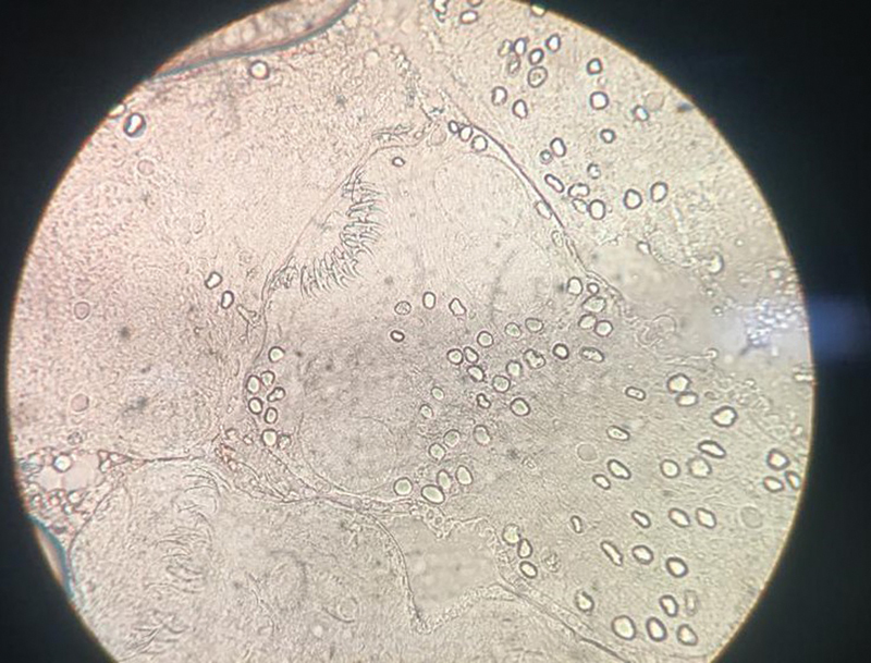
Echinococcosis or hydatid disease is caused by the larval stage of the dog tapeworm, that is, Echinococcus granulosus , E. multilocularis , E. vogeli , or E. oligarthrus . Echinococcus granulosus causes cystic echinococcosis, which has a worldwide distribution. Liver is the most common site, affecting approximately two-third of the patients, whereas lung involvement is seen in approximately 25% of cases. This case is a very rare scenario of bilateral pulmonary hydatid cysts in a young child having exposure to a pet dog with negative hydatid serology and normal eosinophil count.
Keywords: bilateral; echinococcosis; hydatid cyst; pulmonary; serology.
The Indian Association of Laboratory Physicians. This is an open access article published by Thieme under the terms of the Creative Commons Attribution-NonDerivative-NonCommercial License, permitting copying and reproduction so long as the original work is given appropriate credit. Contents may not be used for commercial purposes, or adapted, remixed, transformed or built upon. ( https://creativecommons.org/licenses/by-nc-nd/4.0/ ).
Conflict of interest statement
Conflict of Interest None declared.
Figures

References
-
- Huizinga W KJ, Grant C S, Daar A S. 2nd ed. Oxford, UK: Oxford University Press; 2000. “Hydatid disease.”; pp. 3298–3305.
-
- Erdem C Z, Erdem L O. Radiological characteristics of pulmonary hydatid disease in children: less common radiological appearances. Eur J Radiol. 2003;45(02):123–128. - PubMed
Publication types
LinkOut - more resources
Full Text Sources