Case Reports
. 2022 Sep 2:12:949098.
doi: 10.3389/fonc.2022.949098. eCollection 2022.

Thyroid cancer harboring PTEN and TP53 mutations: A peculiar molecular and clinical case report

Affiliations
Case Reports

Thyroid cancer harboring PTEN and TP53 mutations: A peculiar molecular and clinical case report

Carla Colombo et al. Front Oncol. .

Abstract

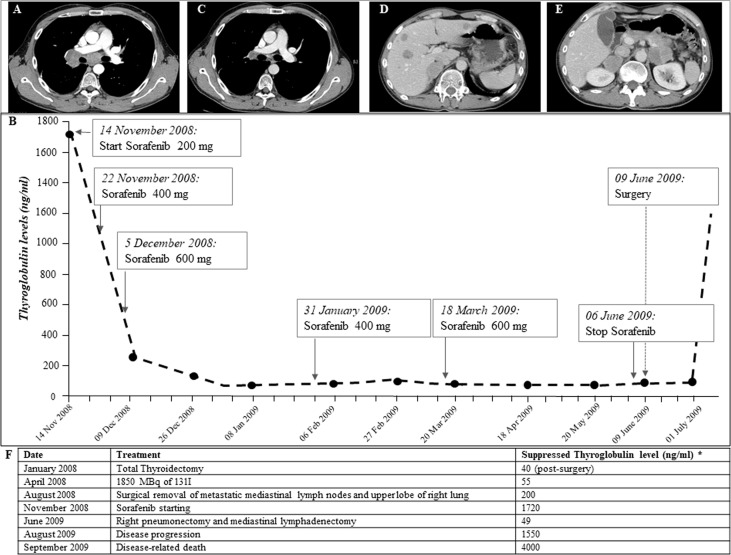
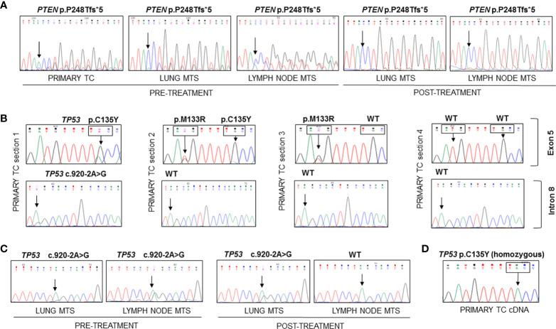
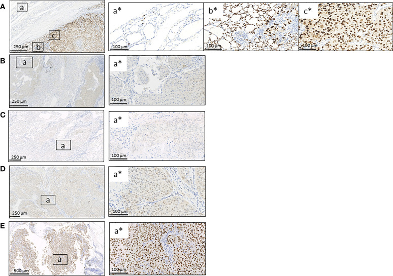
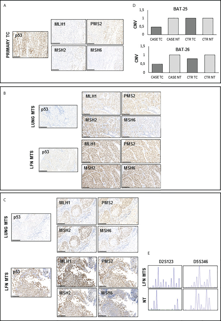
To date, the molecular mechanisms that underline aggressiveness and resistance to tyrosine kinase inhibitors in some thyroid carcinomas (TCs) are not known yet. We report the case of a young patient with a metastatic poorly differentiated (PDTC) and follicular thyroid carcinoma (FTC) refractory to conventional therapies and to Sorafenib. The patient, despite an initial partial response, died of progressive disease 21 months after diagnosis. The genetic analysis performed on the primary tumor and on lymph nodes and distant metastases allowed to identify a frameshift mutation (p.P248Tfs*5) in the PTEN gene, never described in TC. This mutation was present in the primary tumor and, with a lower allelic frequency, in metastases diagnosed after treatment with Sorafenib. Mutations in TP53 (p.C135Y and c.920-2A>G previously detected in anaplastic carcinomas and p.M133R never found in TC) were also detected in the primary tissue together with a mono-allelic expression of the p.C135Y mutant at RNA level. At metastatic sites level, we found only the TP53 splicing mutation c.920-2A>G. The presence of defects in mismatch repair (MMR) proteins and genomic instability was also evaluated. The primary tumor showed a partial expression of MMR proteins together with a strong genomic instability. In conclusion, we demonstrated that the rare combination of somatic PTEN and TP53 mutations in a patient with a metastatic FTC, together with the presence of tumor heterogeneity and genomic instability, might be associated with a high tumor aggressiveness and resistance to treatments.

Keywords: PTEN; Sorafenib; TP53; aggressive follicular thyroid cancer; microsatellite instability; mismatch repair proteins; tyrosine kinase inhibitor.


Conflict of interest statement

The authors declare that the research was conducted in the absence of any commercial or financial relationships that could be construed as a potential conflict of interest.

Figures

Figure 1
Figure 1
(A) CT scan performed on November 2008, before Sorafenib is started. Large lymph node metastases (between 2 and 5.5 cm in diameter) localized in the right hilar region across the pulmonary artery and the main bronchus. (B) Thyroglobulin (Tg) biomarker trend during Sorafenib treatment and after surgical removal of metastases performed after Sorafenib treatment (anti-Tg antibodies persistently negative): Tg values were significantly reduced in the first few weeks of Sorafenib therapy and remained low throughout treatment on different doses. In July 2009, after surgical removal of lymph node and lung metastases, Tg values rose suddenly. (C, D) CT scan performed on July 2009, after following Sorafenib withdrawal and surgical removal of lymph node and lung metastases. Ten liver metastases of 1–2.5 cm size and adrenal metastases (5 cm on the left and 4 cm on the right) were observed. (E) CT scan performed on January 2009, during Sorafenib treatment, showing significant reduction in lymph node metastases (maximum diameter 1 cm). (F) Chronological description of serum suppressed thyroglobulin levels and treatments carried out for thyroid carcinoma.
Figure 2
Figure 2
Sequencing of the PCR amplicons corresponding to exon 7 of PTEN and to exons 5 and 9 of TP53 genes and of RT-PCR amplicon corresponding to exon 5 of TP53 in FFPE and frozen samples. (A) The PTEN p.P248Tfs*5 mutation was found in heterozygosity in the DNA extracted from FFPE primary TC and lung and lymph node metastases samples obtained before and after Sorafenib treatment. (B) Different mutational patterns observed for TP53 in four sections of primary TC. The first sample showed the presence of TP53 p.C135Y mutation in exon 5 and c.920-2A>G splicing variant in intron 8, the second harbored two TP53 mutations (p.M133R and p.C135Y), the third had only the p.M133R mutant, while the last had no mutations. (C) The c.920-2A>G splicing variant was found in all metastases analyzed with the exception of the lymph node metastasis obtained after Sorafenib treatment. (D) The analysis of TP53 transcript encompassing exon 5 showed the presence of the C135Y mutation in homozygous state in the cDNA obtained from frozen primary TC. TC, thyroid cancer; MTS, metastasis.
Figure 3
Figure 3
Immunohistochemistry for p53 protein in FFPE primary TC, contralateral normal thyroid tissue, and lung and lymph node metastases obtained before and after Sorafenib treatment. (A) The immunostaining for p53 was wild type in the contralateral normal thyroid tissue (inset a*) and abnormal/over-expressed with high nuclear expression in primary TC, both FTC/PDCT (inset b*) and PDTC areas (inset c*). (B–D) Lung metastases obtained before (B) and after Sorafenib treatment (D) and lymph node metastasis before TKI (C) showed an abnormal/cytoplasmatic p53 staining with low nuclear expression (for each is shown a selected inset area). (E) The lymph node metastasis obtained after the TKI treatment showed an abnormal/over-expressed p53 pattern with high nuclear expression. FTC, follicular thyroid cancer; PDTC, poorly differentiated thyroid cancer; NT, normal thyroid; TKI, tyrosine kinase inhibitor. Scale bar are shown for each images.
Figure 4
Figure 4
Immunohistochemistry for p53 and DNA mismatch repair (MMR) proteins and microsatellite instability (MSI) detection in available samples before and after Sorafenib treatment (A, C, D) A positive nuclear staining was observed for MHL1, MSH2, MSH6, and PMS2 proteins in all analyzed tissues samples. However, primary TC shows light staining and subclonal loss of expression in all MMR proteins. (B) CNV analysis shows the presence of a LOH for both BAT-25 and BAT-26 loci in the primary TC of our patient. (E) Peak analysis of D2S123 and D5S346 loci, performed using Genemapper 5 software, is clearly differently shaped in the lymph node obtained after Sorafenib treatment with respect to those of and contralateral normal thyroid tissue DNA, indicating a high microsatellite instability. TC, thyroid cancer; MTS, metastasis; NT, normal thyroid; CNV, copy number variation.

Similar articles

Cited by

References

    1. Fugazzola L, Elisei R, Fuhrer D, Jarzab B, Leboulleux S, Newbold K, et al. . European Thyroid association guidelines for the treatment and follow-up of advanced radioiodine-refractory thyroid cancer. Eur Thyroid J (2019) 8:227–45. doi: 10.1159/000502229 - DOI - PMC - PubMed
    1. Kunstman JW, Juhlin CC, Goh G, Brown TC, Stenman A, Healy JM, et al. . Characterization of the mutational landscape of anaplastic thyroid cancer via whole-exome sequencing. Hum Mol Genet (2015) 24:23182329. doi: 10.1093/hmg/ddu749 - DOI - PMC - PubMed
    1. Landa I, Ibrahimpasic T, Boucai L, Sinha R, Knauf JA, Shah RH, et al. . Genomic and transcriptomic hallmarks of poorly differentiated and anaplastic thyroid cancers. J Clin Invest (2016) 126:1052–66. doi: 10.1172/JCI85271 - DOI - PMC - PubMed
    1. Jänicke RU, Graupner V, Budach W, Essmann F. The do's and don'ts of p53 isoforms. Biol Chem (2009) 390:951–63. doi: 10.1515/BC.2009.093 - DOI - PubMed
    1. Shahedian B, Shi Y, Zou M, Farid NR. Thyroid carcinoma is characterized by genomic instability: evidence from p53 mutations. Mol Genet Metab (2001) 72:155–63. doi: 10.1006/mgme.2000.3114 - DOI - PubMed

Publication types