Review
. 2022 Sep 1:13:960702.
doi: 10.3389/fneur.2022.960702. eCollection 2022.

Current status of perimesencephalic non-aneurysmal subarachnoid hemorrhage

Affiliations
Review

Current status of perimesencephalic non-aneurysmal subarachnoid hemorrhage

Kun Hou et al. Front Neurol. .

Abstract

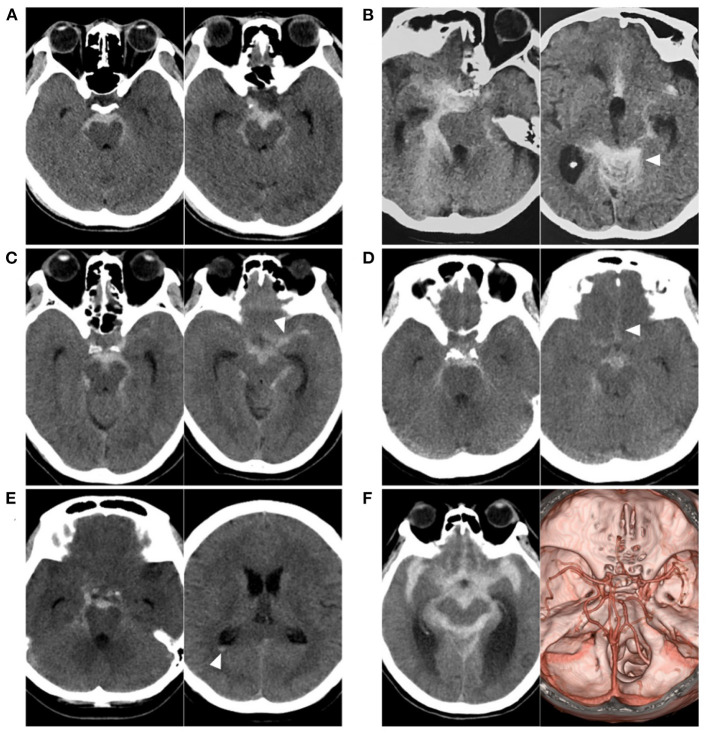
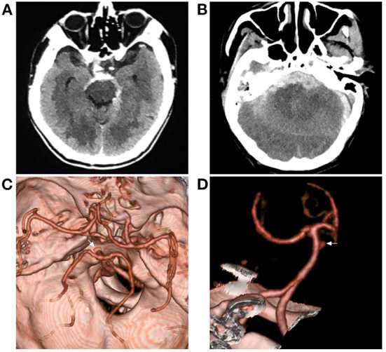
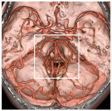
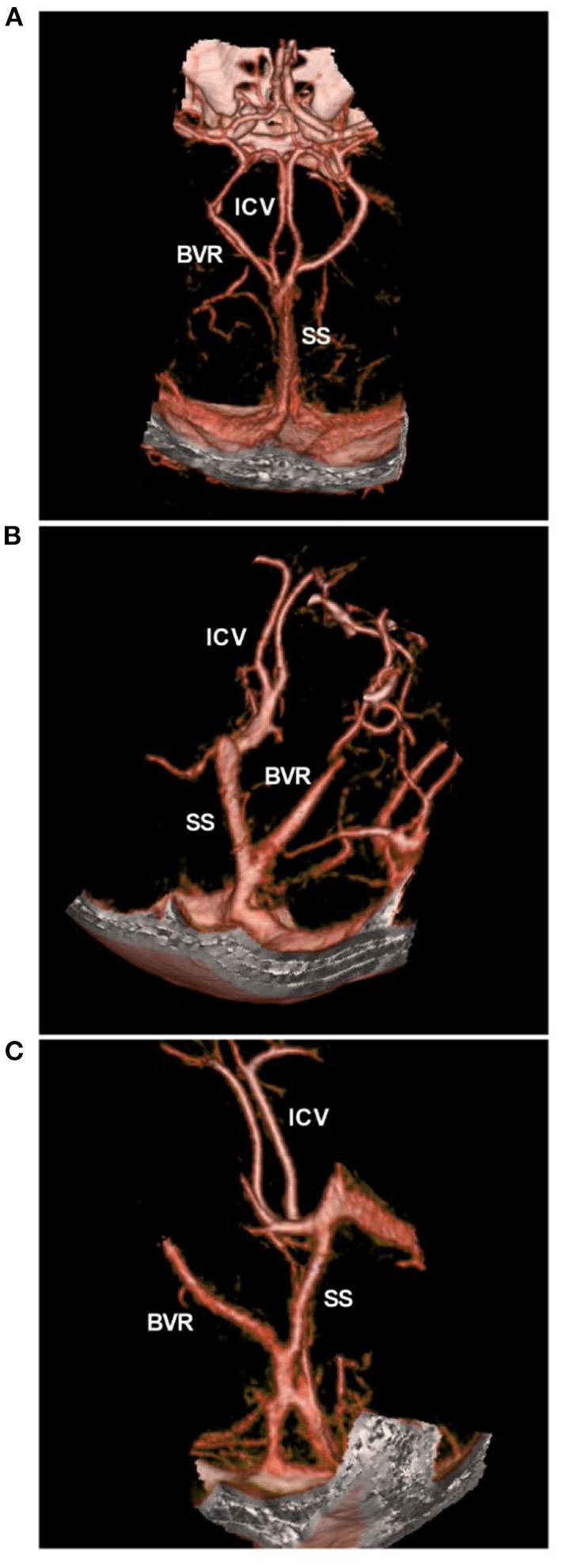
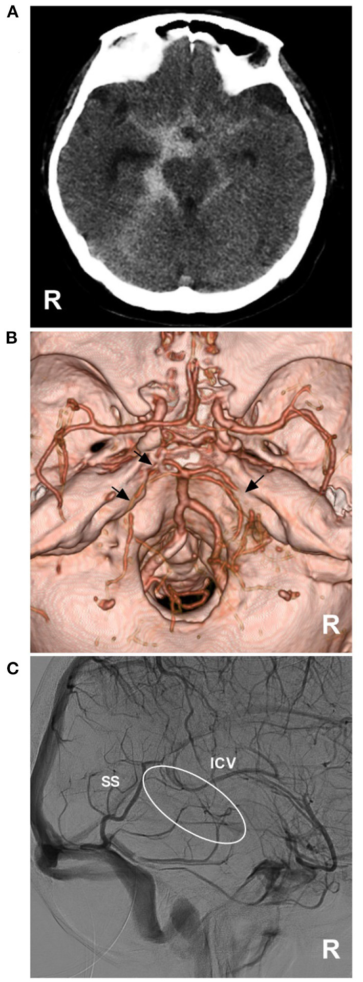
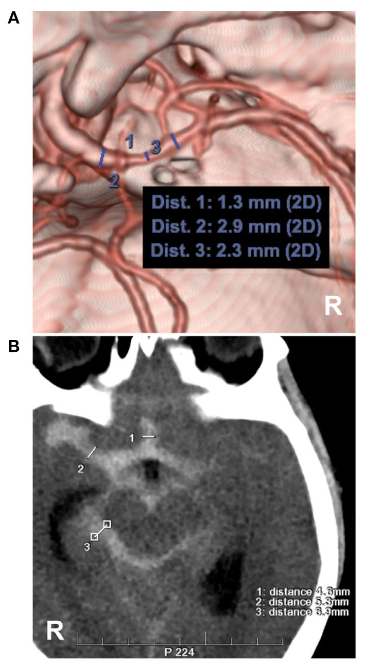
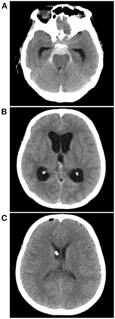
Perimesencephalic nonaneurysmal subarachnoid hemorrhage (PNSAH) is a distinctive disease, representing SAH centered in perimesencephalic cisterns, with negative angiography findings. In recent years, the number of patients with PNSAH has increased significantly; however, the knowledge of PNSAH is insufficient. Therefore, we performed a review of the literature from a PubMed search and recounted our understanding of PNSAH. In this review, we summarized that current high-resolution computed tomography angiography is an acceptable replacement for digital subtraction angiography to rule out aneurysms in PNSAH with strict criteria. The current hypothesis about the etiology of PNSAH is that there is deep vein rupture from aberrant venous anatomy and increased intracranial venous pressure. PNSAH is associated with mild symptoms and lower rates of hydrocephalus and symptomatic vasospasm. For PNSAH, conservative treatment has been the mainstream treatment. PNSAH has a benign clinical course and an excellent prognosis; in long-term follow-up, re-bleeding and death were uncommon.

Keywords: complication; etiology; perimesencephalic non-aneurysmal subarachnoid hemorrhage; prognosis; review.


Conflict of interest statement

The authors declare that the research was conducted in the absence of any commercial or financial relationships that could be construed as a potential conflict of interest.

Figures

Figure 1
Figure 1
Various SAH with negative angiography. (A) CT showing a PNSAH only with focal pre-pontine hemorrhage. (B) CT showing a PNSAH with involvement of quadrigeminal cistern (arrowhead). (C) CT showing a PNSAH extended to the basal part of the Sylvian fissure (arrowhead). (D) CT showing a PNSAH extended to the posterior part of the interhemispheric fissure (arrowhead). (E) CT showing a PNSAH with intraventricular sedimented blood (arrowhead). (F) Left: CT showing a diffuse non-PNSAH; Right: the intracranial aneurysm was not found on CTA; the drainage pattern of the deep vein around vein of Galen was normal. CT, computed tomography; CTA, computed tomography angiography; PNSAH, perimesencephalic non-aneurysmal subarachnoid hemorrhage; SAH, subarachnoid hemorrhage.
Figure 2
Figure 2
Aneurysmal SAH with perimesencephalic bleeding pattern. (A) Immediate CT of the onset showing SAH with typical perimesencephalic bleeding pattern. (B) Repeated CT 6 h after the onset showing a second SAH with the thick hemorrhage in front of the brainstem. (C,D) CTA showing an aneurysm of the basilar artery (arrows). CT, computed tomography; CTA, computed tomography angiography; SAH, subarachnoid hemorrhage.
Figure 3
Figure 3
Region of possible etiology of PNSAH. By extending the time of image acquisition of computed tomography angiography, both arteries and deep veins are detected simultaneously. The possible etiology of PNSAH should focus on the frame region. PNSAH, perimesencephalic non-aneurysmal subarachnoid hemorrhage.
Figure 4
Figure 4
BVR variants in CTA. (A) CTA showing the normal pattern of deep veins, bilateral ICVs and BVRs that flow into the vein of Galen. (B,C) CTA showing the BVR that flowed into the straight sinus. BVR, Basal vein of Rosenthal; CTA, computed tomography angiography; ICV, internal cerebral vein, SS, straight sinus.
Figure 5
Figure 5
PNSAH with BVR absence. (A) CT showing a PNSAH that extended to the posterior part of the anterior interhemispheric fissure and the basal part of the Sylvian fissure. (B) CTA showing the multiple vasospasms (arrows) of the bilateral posterior cerebral arteries, without positive findings of bleeding. (C) DSA showing the absence of the right BVR (circle). BVR, Basal vein of Rosenthal; CT, computed tomography; CTA, computed tomography angiography; DSA, digital subtraction angiography; ICV, internal cerebral vein; PNSAH, perimesencephalic non-aneurysmal subarachnoid hemorrhage; R, right; SS, straight sinus.
Figure 6
Figure 6
Measurements of intracranial vasospasm and SAH thickness. (A) CTA showing the diameter measurement of the MCA. No. 1, 2 and 3 showing the diameters of the different locations of the MCA, the location of No. 1 had a moderate vasospasm (33–66%, 1.3/2.3 and 1.3/2.9). (B) CT showing the measurements of the Barrow Neurological Institute scale, No. 1, 2 and 3 showing the subarachnoid hemorrhage thickness (4.6, 5.3, 6.9 mm), by the measurement across the thickest-appearing regions of the cistern or fissure. CT, computed tomography; CTA, computed tomography angiography; MCA, middle cerebral artery; R, right.
Figure 7
Figure 7
External ventricle drainage in a PNSAH. (A) CT showing a classic PNSAH. (B) CT showing acute lateral ventricle dilation. (C) CT showing the normal size of lateral ventricle after external ventricle drainage. CT, computed tomography; PNSAH, perimesencephalic non-aneurysmal subarachnoid hemorrhage.

References

    1. Andaluz N, Zuccarello M. Yield of further diagnostic work-up of cryptogenic subarachnoid hemorrhage based on bleeding patterns on computed tomographic scans. Neurosurgery. (2008) 62:1040–6. 10.1227/01.neu.0000325865.22011.1f - DOI - PubMed
    1. Gama Lobo G, Fragata I. Long-term global and focal cerebral atrophy in perimesencephalic subarachnoid hemorrhage-a case-control study. Neuroradiology. (2022) 64:669–74. 10.1007/s00234-021-02804-w - DOI - PubMed
    1. Nguyen I, Caton MT, Tonetti D, Abla A, Kim A, Smith W, et al. . Angiographically occult subarachnoid hemorrhage: yield of repeat angiography, influence of initial CT bleed pattern, and sources of diagnostic error in 242 consecutive patients. AJNR Am J Neuroradiol. (2022). 10.3174/ajnr.A7483 - DOI - PMC - PubMed
    1. van Gijn J, van Dongen KJ, Vermeulen M, Hijdra A. Perimesencephalic hemorrhage: a nonaneurysmal and benign form of subarachnoid hemorrhage. Neurology. (1985) 35:493–7. 10.1212/WNL.35.4.493 - DOI - PubMed
    1. Mensing LA, Vergouwen MDI, Laban KG, Ruigrok YM, Velthuis BK, Algra A, et al. . Perimesencephalic hemorrhage: a review of epidemiology, risk factors, presumed cause, clinical course, and outcome. Stroke. (2018) 49:1363–70. 10.1161/STROKEAHA.117.019843 - DOI - PubMed

LinkOut - more resources