. 2022 Aug 15;12(8):3601-3624.
eCollection 2022.

Cordycepin enhances radiosensitivity to induce apoptosis through cell cycle arrest, caspase pathway and ER stress in MA-10 mouse Leydig tumor cells

Affiliations

Cordycepin enhances radiosensitivity to induce apoptosis through cell cycle arrest, caspase pathway and ER stress in MA-10 mouse Leydig tumor cells

Yi-Ping Lee et al. Am J Cancer Res. .

Abstract

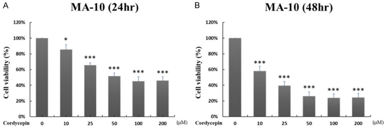
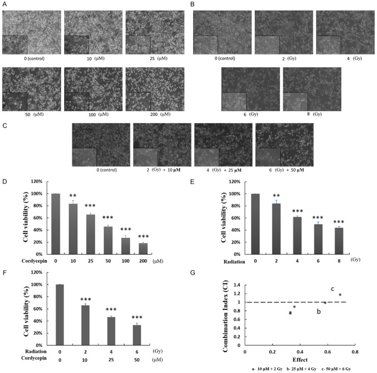
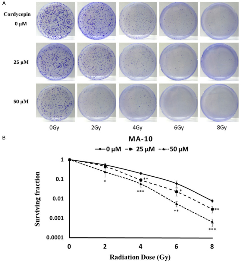
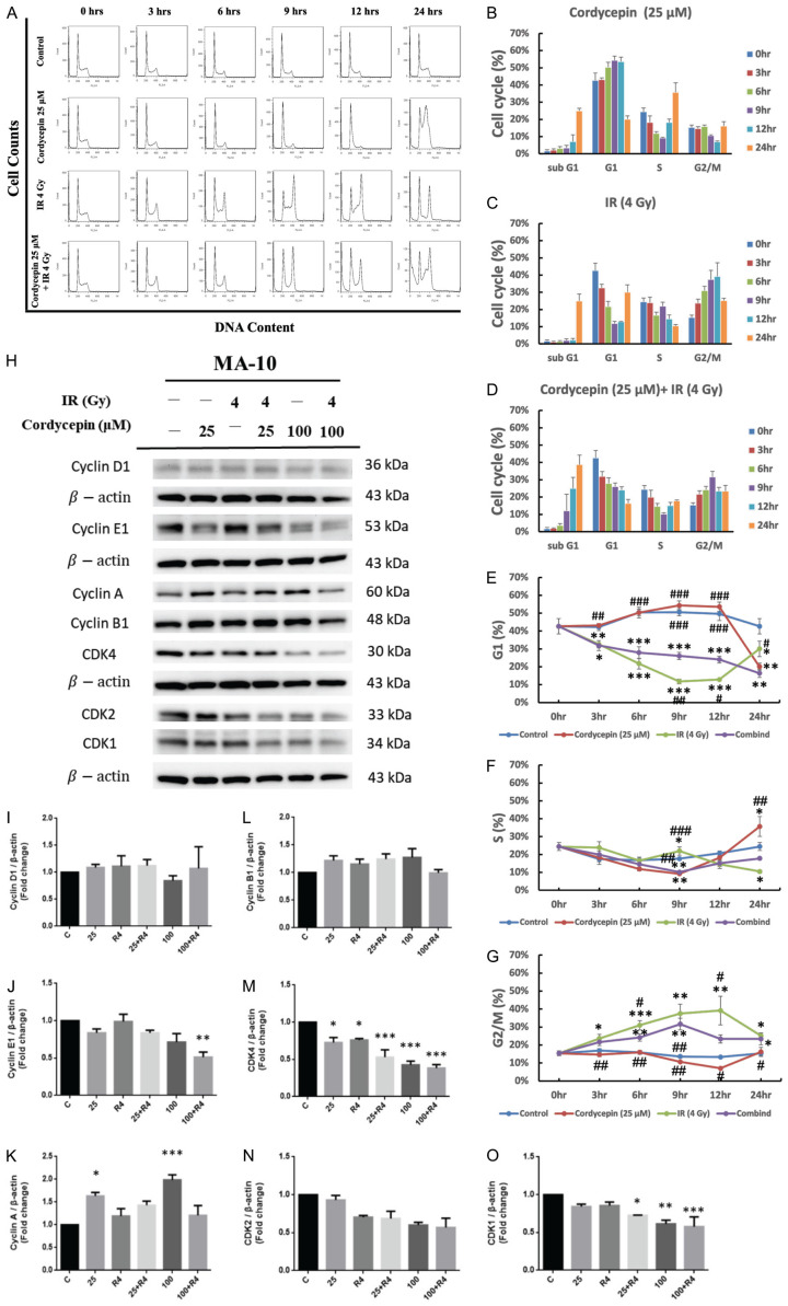
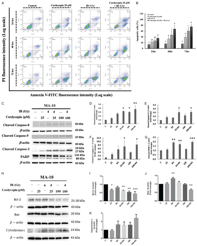
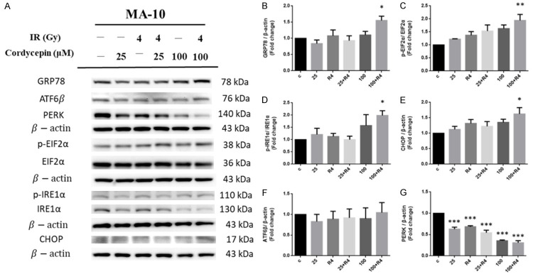
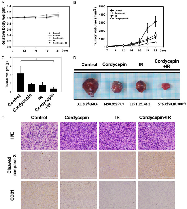
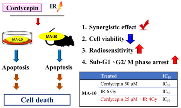
Radiotherapy is a localized treatment commonly used in various types of cancer. However, major limitation of radiotherapy is the development of resistance of tumor cells to radiosensitivity. Cordycepin, a predominant functional component of the Cordyceps sinensis, is considered to use in treating tumor cells. In the present study, we investigated the anticancer effect of the combination of radiation and cordycepin in the treatment of Leydig tumor cells. Results showed that the combination treatment has a synergistic effect significantly suppress cell viability and enhance the radiosensitivity in MA-10 mouse Leydig tumor cells. The combination treatment induced MA-10 cell apoptosis through increasing levels of cleaved caspase-3/-8/-9, poly ADP-ribose polymerase (PARP), and cytochrome c and decreasing levels of B-cell lymphoma 2 (Bcl-2). In addition, prolonged sub-G1 and G2/M arrest accompany with cell cycle-related protein regulation was observed in cells that received the combination treatment. The endoplasmic reticulum (ER) stress-related protein expressions were regulated after MA-10 cells treating with a combination of 100 μM cordycepin and 4 Gy radiation. Furthermore, the combination treatment also decreased the Leydig tumor mass by increasing cell apoptosis in tumor-bearing mice. In conclusion, cordycepin enhances radiosensitivity to induce mouse Leydig tumor cells toward apoptosis in vitro and in vivo. This study will provide a scientific basis for the development of therapeutic regimen of testicular cancer.

Keywords: Cordycepin; ER stress; Leydig tumor cell; MA-10 cell; apoptosis; caspase; cell cycle; radiation.


Conflict of interest statement

None.

Figures

Figure 1
Figure 1
Cordycepin reduced MA-10 growth ability in time- and dose-dependent manners. MA-10 cells were treated with 10, 25, 50, 100 and 200 μM cordycepin for 24 hr (A), and for 48 hr (B), respectively. Cell viabilities were examined by MTT viability test. Results were presented as percentages of cell growth of each group relative to control group. *P<0.05, and ***P<0.001 indicate significant statistical difference compared to the control group.
Figure 2
Figure 2
Cordycepin, radiation, and the combination treatment decreased MA-10 cell viability in dose-dependent manners. MA-10 cells were treated with 0, 10, 25, 50, 100 and 200 μM cordycepin for 24 hr (A, D), with 0, 2, 4, 6 and 8 Gy radiation for 24 hr (B, E), and with the combination treatments of cordycepin and radiation for 24 hr (C, F), respectively. The morphology of cells was examined using light microscope and images were captured by digital camera (A-C). Cell viabilities were examined by Trypan blue exclusion test (D-F). Results were presented as percentages of cell growth relative to control groups. CalcuSyn software analysis was used to determine the synergistic effects of combination treatment of cordycepin and radiation on MA-10 cells (G). **P<0.01, and ***P<0.001 indicate significant statistical difference compared to the control group. The data points at “a” and “b” indicate the synergies.
Figure 3
Figure 3
Cordycepin enhanced radiosensitivity in MA-10 cells. MA-10 cells were treated with radiation (0, 2, 4, 6 and 8 Gy) combined with different concentrations of cordycepin (0, 25 and 50 μM). Cells were plated in 6 cm dishes for 8 days. Dishes were stained with crystal violet (A). Colonies containing >50 cells were scored as positive and then the radiation dose-response survival curves were determined (B). The data represent the mean ± S.D. *P<0.05, **P<0.01, and ***P<0.001 indicate significant statistical difference compared to the control group.
Figure 4
Figure 4
Cordycepin, radiation, and the combination treatment regulated cell cycle distribution in MA-10 cells. MA-10 cells were treated without or with 25 μM cordycepin and 4 Gy radiation for 3, 6, 9, 12 and 24 hr, respectively. Cells were fixed and then stained with propidium iodide (PI), and cell cycle was measured by flow cytometry. The distribution and percentage of cells in sub G1, G1, S and G2/M phase of the cell cycle are illustrated (A-G). MA-10 cells were treated without or with different concentrations of cordycepin (25 and 100 µM) and radiation (4 Gy) for 24 hr, respectively. Cyclin D1, Cyclin E1, Cyclin A, Cyclin B1, CDK4, CDK2 and CDK1 were detected by western blotting (H-O). *P<0.05, **P<0.01, and ***P<0.001 indicate significant statistical difference compared to the control group. #P<0.05, ##P<0.01 and ###P<0.001 indicate significant statistical difference compared to the combination treatment group.
Figure 5
Figure 5
Cordycepin and radiation induced cell apoptosis in MA-10 cells. MA-10 cells were treated without or with 25 or 50 μM cordycepin and/or 4 Gy radiation for 24, 48 and 72 hr, respectively, and stained with annexin V and propidium iodide (PI). The original density plots of Flow Cytometry analysis were showed (A). Data were analyzed by two-way ANOVA with Tukey’s multiple comparisons test. Different superscripts above each column indicated significant difference among each treatment (P<0.05) (B). Apoptosis related protein were detected by western blotting and normalized with β-actin (43 kDa) in each lane (C-K). *P<0.05, **P<0.01, and ***P<0.001 indicate significant statistical difference compared to the control group.
Figure 6
Figure 6
The effect of cordycepin and radiation on ER stress pathway in MA-10 cells. MA-10 cells were treated without or with different concentrations of cordycepin (25 and 100 µM) and/or radiation (4 Gy) for 24 hr, respectively. GRP78, p-EIF2α, p-IRE1α, CHOP, ATF6β and PERK were detected by western blotting (A). The integrated optical densities (IOD) of GRP78, p-EIF2α, p-IRE1α, CHOP, ATF6β and PERK were normalized with β-actin (43 kDa) in each lane. *P<0.05, **P<0.01, and ***P<0.001 indicate significant statistical difference compared to the control group (B-G).
Figure 7
Figure 7
Combination treatment synergistically inhibits tumorigenesis of MA-10 cells in tumor-bearing C57BL/6 mice. C57BL/6 mice were treated with IR (4 Gy) or cordycepin (20 mg/kg) alone or in combination. A. Measurement of body weight in mice 2-3 days per week. B. MA-10 transplanted tumor growth curves in mice. Data are presented as the relative tumor volume normalized to the initial tumor volume measured on Day 7 as a function of time after start of treatment. C. Measurement of tumor weight of transplanted MA-10 in mice. D. Direct observation of mice with tumors from the control and cordycepin, IR alone or in combination groups. E. H&E staining and immunohistochemical staining for analysis of Cleaved caspase 3-positive and CD31-positive cells (brown), Microscopic view with 200X. *P<0.05 indicates significant statistical difference compared to the control group.
Figure 8
Figure 8
The effect of the combination treatment of cordycepin and radiation in mouse Leydig tumor cells. The combination treatment of cordycepin and radiation has a synergistic effect could significantly suppress cell viability and enhance the radiosensitivity in MA-10 cells. In addition, cordycepin and radiation could significantly induce sub-G1 and G2/M phase arrest in MA-10 cells. Moreover, the combination treatment of cordycepin and radiation induces MA-10 cells to progress toward apoptosis in vitro and in vivo.

Similar articles

Cited by

References

    1. Milose JC, Filson CP, Weizer AZ, Hafez KS, Montgomery JS. Role of biochemical markers in testicular cancer: diagnosis, staging, and surveillance. Open Access J Urol. 2011;4:1–8. - PMC - PubMed
    1. Papworth DG, Lloyd RA. Cancer survival in the USA, 1973-1990: a statistical analysis. Br J Cancer. 1998;78:1514–1515. - PMC - PubMed
    1. Richiardi L, Scelo G, Boffetta P, Hemminki K, Pukkala E, Olsen JH, Weiderpass E, Tracey E, Brewster DH, McBride ML, Kliewer EV, Tonita JM, Pompe-Kirn V, Kee-Seng C, Jonasson JG, Martos C, Brennan P. Second malignancies among survivors of germ-cell testicular cancer: a pooled analysis between 13 cancer registries. Int J Cancer. 2007;120:623–631. - PubMed
    1. Pan BS, Wang YK, Lai MS, Mu YF, Huang BM. Cordycepin induced MA-10 mouse Leydig tumor cell apoptosis by regulating p38 MAPKs and PI3K/AKT signaling pathways. Sci Rep. 2015;5:13372. - PMC - PubMed
    1. Petersen PM, Skakkebaek NE, Rorth M, Giwercman A. Semen quality and reproductive hormones before and after orchiectomy in men with testicular cancer. J Urol. 1999;161:822–826. - PubMed

LinkOut - more resources