

Clinical Clues to Differentiate between Dermatophyte Onychomycosis (DP-OM) and Dermatophytoma-Like Traumatic Onychodystrophy (DP-TO)
- PMID: 36119939
- PMCID: PMC9481322
- DOI: 10.1155/2022/8519376
Clinical Clues to Differentiate between Dermatophyte Onychomycosis (DP-OM) and Dermatophytoma-Like Traumatic Onychodystrophy (DP-TO)
Erratum in
-
Corrigendum to "Clinical Clues to Differentiate Between Dermatophyte Onychomycosis (DP-OM) and Dermatophytoma-Like Traumatic Onychodystrophy (DP-TO)".Biomed Res Int. 2025 Jul 28;2025:9862803. doi: 10.1155/bmri/9862803. eCollection 2025. Biomed Res Int. 2025. PMID: 40761506 Free PMC article.
Abstract
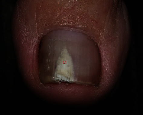
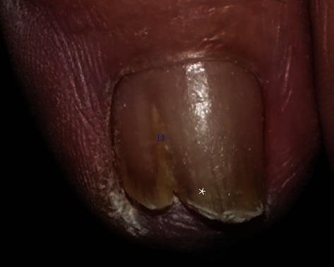
Background: Dermatophytoma is a recalcitrant condition of onychomycosis (OM). It presents as a white- or yellow-colored fungal mass that appears linear/triangular or round on a nail plate. Traumatic onychodystrophy (TO) can present with dermatophytoma-like lesions. Typically, OM and TO are not clinically distinguishable. Mycological testing is the gold standard for differentiating these disorders.
Objectives: This study is aimed at differentiating between the clinical and dermoscopic factors related to dermatophytoma onychomycosis (DP-OM) and dermatophytoma-like traumatic onychodystrophy (DP-TO).
Methods: A retrospective study was conducted of patients with dermatophytoma-like nail lesions who visited the Siriraj Nail Clinic between January 2010 and July 2020. The diagnosis of DP-OM was made by direct microscopy, fungal cultures, and histopathology of nail clippings.
Results: A total of 36 nails were included in the study. Thirteen nails were DP-OM, and 23 nails were DP-TO. The demographic data and risk factors for the 2 groups were not significantly different. Dermatophytoma lesions were found on the lateral side of nails in 12 cases of DP-OM (92.3%) and 11 cases of DP-TO (47.8%; P = 0.008). DP-OM was associated with longitudinal striae adjacent to dermatophytoma (69.2% vs. 30.4%; P = 0.024), sulfur-nugget-like subungual debris (23.1% vs. 0%; P = 0.040), and scale on the ipsilateral foot (69.2% vs. 8.7%; P < 0.001). DP-TO was associated with a homogenous, whitish discoloration (47.8% vs. 7.7%; P = 0.014) and a sharp edge of the onycholytic area (43.5% vs. 0%; P = 0.005).
Conclusions: The lateral location of dermatophytoma, adjacent striae, sulfur-nugget-like debris, and scale on the ipsilateral foot were significantly associated with DP-OM. Dermoscopic examination (dorsal and hyponychium views) and foot examination are beneficial for distinguishing between DP-OM and DP-TO.
Copyright © 2022 Sumanas Bunyaratavej et al.
Conflict of interest statement
The authors have no conflicts of interest to declare.
Figures
References
MeSH terms
Substances
LinkOut - more resources
Full Text Sources
Medical
