. 2021 Jul 15;14(1):70-78.
doi: 10.1016/j.chmed.2021.07.002. eCollection 2022 Jan.

Didymin attenuates doxorubicin-induced cardiotoxicity by inhibiting oxidative stress

Affiliations

Didymin attenuates doxorubicin-induced cardiotoxicity by inhibiting oxidative stress

Rongchang Chen et al. Chin Herb Med. .

Abstract

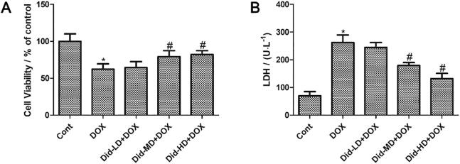
Objective: This study was designed to investigate the protective effects of didymin (Did) on doxorubicin (DOX)-induced cardiotoxicity.

Methods: After pretreatment with Did (2, 4, 8 mg/kg intraperitoneal i.p.) for 7 d, the male C57 mice were injected with single dose of DOX (20 mg/kg i.p.). The cardioprotective effect of Did was observed on the 7th day after DOX treatment.

Results: DOX delayed body growth and caused cardiac tissue injury, oxidative stress, and mitochondrial dysfunction. Similar experiments in H9C2 cardiomyocytes showed that DOX reduced cell viability, increased generation of reactive oxygen species (ROS) and fragmentation of DNA, decreased mitochondrial membrane potential, and induced cardiomyocyte apoptosis. However, all of these adverse effects were suppressed by Did pretreatment. Did increased protein expression of glutamate-L-cysteine ligase catalytic subunit (GCL), heme oxygenase 1 (HO-1), and nuclear factor erythroid 2-related factor 2 (Nrf2). Besides, Did also induced activation of PI3K/AKT.

Conclusion: These findings indicated Did prevented DOX-induced cardiac injury and apoptosis via activating PI3K/AKT/Nrf2 signaling pathway.

Keywords: Nrf2; PI3K/Akt; cardiotoxicity; didymin; doxorubicin.


Conflict of interest statement

The authors declare that they have no known competing financial interests or personal relationships that could have appeared to influence the work reported in this paper.

Figures

Fig. 1
Fig. 1
Effects of Did on DOX-induced myocardial injury in vivo (mean ± SD, n = 10). Mice were treated with vehicle or DOX with or without Did pretreatment. At day 7, body weight of mice was determined (A); Effects of Did and DOX on AST (B), LDH (C), and CK (D) activities were measured. *P < 0.05 vs control group, and #P < 0.05 vs DOX group. Did-LD + DOX: low-dose Did group (2 mg/kg); Did-MD + DOX: medium-dose Did group (4 mg/kg); Did-HD + DOX: high-dose Did group (8 mg/kg).
Fig. 2
Fig. 2
Effects of Did and DOX on cardiac function. (A) Representative M−mode ECG images are shown. Echocardiography values are expressed as mean ± SD. (B) EF, ejection fraction. (C) FS, fractional shortening. Results are represented as the mean ± SE (n = 6). *P < 0.05 vs control group, and #P < 0.05 vs DOX group. Did-LD + DOX: low-dose Did group (2 mg/kg); Did-MD + DOX: medium-dose Did group (4 mg/kg); Did-HD + DOX: high-dose Did group (8 mg/kg).
Fig. 3
Fig. 3
Effects of Did and DOX on histological changes in mice hearts, as shown by HE staining (A) under electron microscope. Effects of Did and DOX on ultra structure changes in mice hearts observed under transmission electron microscopy (B). Did-LD + DOX: low-dose Did group (2 mg/kg); Did-MD + DOX: medium-dose Did group (4 mg/kg); Did-HD + DOX: high-dose Did group (8 mg/kg).
Fig. 4
Fig. 4
Effects of Did and DOX on SOD (A), CAT (B), GSH-px (C), and MDA (D) activities in heart tissues of mice (mean ± SD, n = 10). *P < 0.05 vs control group, and #P < 0.05 vs DOX group. Did-LD + DOX: low-dose Did group (2 mg/kg); Did-MD + DOX: medium-dose Did group (4 mg/kg); Did-HD + DOX: high-dose Did group (8 mg/kg).
Fig. 5
Fig. 5
Effects of Did and DOX on cell viability (A) and LDH release (B) in vitro. H9c2 cells were treated with different concentrations of Did for 4 h, and further exposed to 1 µmol/L DOX for 24 h (mean ± SD, n = 8).. *P < 0.05 vs control group, and #P < 0.05 vs DOX group. Did-LD + DOX: low-dose Did group (5 μg/ml); Did-MD + DOX: medium-dose Did group (10 μg/ml); Did-HD + DOX: high-dose Did group (20 μg/ml).
Fig. 6
Fig. 6
Effects of Did and DOX on apoptosis in vitro (mean ± SD, n = 8). (A) Hoechst 33342 staining was used to measure the apoptosis in H9c2 cells. (B) Cells stained with JC-1 dye were visualized by fluorescence microscopy. Quantitative analysis of Hoechst 33342 (C) and JC-1 (D) staining was evaluated. *P < 0.05 vs control group, and #P < 0.05 vs DOX group. Did-LD + DOX: low-dose Did group (5 μg/ml); Did-MD + DOX: medium-dose Did group (10 μg/ml); Did-HD + DOX: high-dose Did group (20 μg/ml).
Fig. 7
Fig. 7
ROS levels in different groups by fluorescence staining (mean ± SD, n = 8). *P < 0.05 vs control group, and #P < 0.05 vs DOX group. Did-LD + DOX: low-dose Did group (5 μg/mL); Did-MD + DOX: medium-dose Did group (10 μg/mL); Did-HD + DOX: high-dose Did group (20 μg/mL).
Fig. 8
Fig. 8
Effects of Did and DOX on SOD (A), MDA (B), CAT (C), and GSH-Px (D) activities and HO-1 (E), and GCL (F) levels in H9c2 (mean ± SD, n = 8). *P < 0.05 vs control group, and #P < 0.05 vs DOX group. Did-LD + DOX: low-dose Did group (5 μg/mL); Did-MD + DOX: medium-dose Did group (10 μg/mL); Did-HD + DOX: high-dose Did group (20 μg/mL).
Fig. 9
Fig. 9
Protein levels of Bax, Bcl-2, Caspase-3 and Caspase-9 in H9c2 cells determined using (A) Western blot analysis and (B) expressed as the fold changes over the control. The protein levels of phosphorylated and total AKT, PI3K, Nrf2, HO-1, and GCL were measured using (C) Western blot analysis and (D) were expressed as the fold changes over the control. Results are represented as the mean ± SD (n = 8). *P < 0.05 vs control group, and #P < 0.05 vs DOX group.

References

    1. Abdullah C.S., Alam S., Aishwarya R., Miriyala S., Bhuiyan M.A.N., Panchatcharam M.…Bhuiyan M.S. Doxorubicin-induced cardiomyopathy associated with inhibition of autophagic degradation process and defects in mitochondrial respiration. Scientific Reports. 2019;9:2002. - PMC - PubMed
    1. Benjanuwattra J., Siri-Angkul N., Chattipakorn S.C., Chattipakorn N. Doxorubicin and its proarrhythmic effects: A comprehensive review of the evidence from experimental and clinical studies. Pharmacological Research. 2020;151 - PubMed
    1. Catanzaro M.P., Weiner A., Kaminaris A., Li C., Cai F., Zhao F.…Liang Q. Doxorubicin-induced cardiomyocyte death is mediated by unchecked mitochondrial fission and mitophagy. FASEB Journal: Official Publication of the Federation of American Societies for Experimental Biology. 2019;33:11096–11108. - PMC - PubMed
    1. Chen R., Sun G., Yang L., Wang J., Sun X. Salvianolic acid B protects against doxorubicin induced cardiac dysfunction via inhibition of ER stress mediated cardiomyocyte apoptosis. Toxicology Research. 2016;5:1335–1345. - PMC - PubMed
    1. Chen, R.C., Xu, X.D., Liu, X. Z., Sun, G.B., Zhu, Y.D., Dong, X., Wang, J., Zhang, H.J., Zhang, Q., Sun, X.B. (2015). Total flavonoids from Clinopodium chinense (Benth.) O. Ktze protect against doxorubicin-induced cardiotoxicity in vitro and in vivo. Evidence-based Complementary and Alternative Medicine: Ecam, 2015: 472565. - PMC - PubMed

LinkOut - more resources