. 2022 Aug 30:13:952337.
doi: 10.3389/fphar.2022.952337. eCollection 2022.

Protective effects of menthol against sepsis-induced hepatic injury: Role of mediators of hepatic inflammation, apoptosis, and regeneration

Affiliations

Protective effects of menthol against sepsis-induced hepatic injury: Role of mediators of hepatic inflammation, apoptosis, and regeneration

Asmaa I Matouk et al. Front Pharmacol. .

Abstract

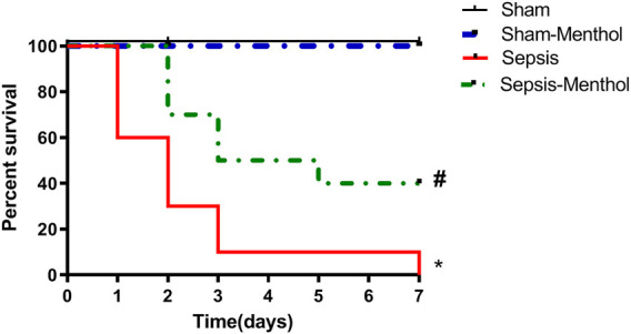
Liver dysfunction in sepsis is a major complication that amplifies multiple organ failure and increases the risk of death. Inflammation and oxidative stress are the main mediators in the pathophysiology of sepsis. Therefore, we investigated the role of menthol, a natural antioxidant, against sepsis-induced liver injury in female Wistar rats. Sepsis was induced by cecal ligation and puncture (CLP). Menthol (100 mg/kg) was given intragastric 2 h after CLP. Blood samples and liver tissues were collected 24 h after surgery. Menthol significantly (p < 0.05) attenuated the sepsis-induced elevation in serum liver enzymes and improved the hepatic histopathological changes. Menthol treatment significantly (p < 0.05) decreased hepatic levels of tumor necrosis factor-alpha, malondialdehyde, total nitrite, and cleaved caspase-3. It restored the hepatic levels of superoxide dismutase and reduced glutathione. Additionally, menthol significantly (p < 0.05) increased hepatic levels of B-cell lymphoma 2 (Bcl-2); an anti-apoptotic factor, and proliferating cell nuclear antigen (PCNA), a biomarker of regeneration and survival. Our results showed the therapeutic potential of menthol against liver injury induced by sepsis.

Keywords: PCNA; apoptosis; clp; hepatoprotection; tNF-alpha.


Conflict of interest statement

The authors declare that the research was conducted in the absence of any commercial or financial relationships that could be construed as a potential conflict of interest.

Figures

FIGURE 1
FIGURE 1
Effect of menthol on CLP-induced mortality. Induction of sepsis by CLP model resulted in 0% survival at the end of the 7th day. Treatment with menthol (100 mg/kg, I. G) 2 h after CLP improved the survival by 40% at the end of the 7th day. The sham group and sham-menthol (100 mg/kg, I. G) group had no mortality throughout the study. Data are presented as a percentage of survival of rats (n = 10 per group). * significant difference from the sham group at p ˂0.05. # significant difference from the sepsis group at p ˂0.05.
FIGURE 2
FIGURE 2
The effect of menthol (100 mg/kg, I.G) on serum levels of ALT (Figure 2A) and AST (Figure 2B) in CLP septic rats. Figure 2C: Photomicrographs showing liver sections from all groups (n = 6, each) examined by H&E-stained (200x). The sham groups [sham and sham-menthol; (A) and (B)] showed normal liver cells (arrow head) with normal central vein (arrow). The liver tissue from the sepsis group (C) showed disrupted hepatic cells architecture (arrow head) with dilated central veins (arrow). Liver tissues from the menthol-treated septic rats (D) showed normal liver cells. Figure 2D: Scoring the histopathological changes; 0: absent, 1: <25%, 2: >25% and <50%, 3: >50% and <75% and 4: >75% of the entire section showed histopathological alterations. Data represented as a mean score of each group for each observed histopathological alteration. * significant difference from the sham group at p ˂0.05. # significant difference from the sepsis group at p ˂0.05.
FIGURE 3
FIGURE 3
The effect of menthol (100 mg/kg, I.G) on hepatic TNF-α in CLP sepsis model; (A); Representative photomicrographs showing TNF-α immunoreactivity in liver tissue, (B); Bar charts showing semi-quantitative analysis of data in A from sections of the sham, sham-menthol, sepsis, and sepsis-menthol groups. * significant difference from the sham group at p ˂0.05. # significant difference from the sepsis group at p ˂0.05. Data represented as mean ± S.E (n = 6).
FIGURE 4
FIGURE 4
The effect of menthol (100 mg/kg, I.G) on hepatic cleaved caspase-3 and Bcl-2 levels in CLP sepsis model. (A); Representative photomicrographs showing cleaved caspase-3 immunoreactivity in liver tissues. (B); Bar charts showing semi-quantitative analysis of data in A from tissue sections of the sham, sham-menthol, sepsis, and sepsis-menthol groups. (C) Representative photomicrographs showing Bcl-2 immunoreactivity in liver tissues. (D) Bar charts showing semi-quantitative analysis of data in C from tissue sections of the sham, sham-menthol, sepsis, and sepsis-menthol groups * significant difference from the sham group at p ˂0.05. # significant difference from the sepsis group at p ˂0.05. Data represented as mean ± S.E (n = 6).
FIGURE 5
FIGURE 5
The effect of menthol (100 mg/kg, I.G) on hepatic PCNA levels in CLP sepsis model (A); Representative photomicrographs showing PCNA immunoreactivity in liver tissues. (B); Bar charts showing semi-quantitative analysis of the data from all groups. * significant difference from the sham group at p ˂0.05. # significant difference from the sepsis group at p ˂0.05. Data represented as mean ± S.E (n = 6).
FIGURE 6
FIGURE 6
Correlation matrix for different parameters included in the study. Pearson correlation coefficient (r) was used to measure the correlation. If r is between |0.3| and |0.7|, a moderate correlation is indicated. If r >|0.7|, a strong correlation is indicated. If r<|0.3|, a weak correlation is indicated. Positive values indicate a positive correlation, while negative values indicate a negative correlation. The color of the scale bar to the right ranges from violet to red, indicating r values from +1 to -1. The graph is colored according to the scale. TNF-α: tumor necrosis factor-alpha; PCNA: proliferating cell nuclear antigen; Bcl-2: B-cell lymphoma 2.

References

    1. Abcejo A., Andrejko K. M., Ochroch E. A., Raj N. R., Deutschman C. S. (2011). Impaired hepatocellular regeneration in murine sepsis is dependent on regulatory protein levels. Shock 36 (5), 471–477. 10.1097/SHK.0b013e31822d60ff - DOI - PMC - PubMed
    1. Aboyoussef A. M., Mohammad M. K., Abo-Saif A. A., Messiha B. A. S. (2021). Granisetron attenuates liver injury and inflammation in a rat model of cecal ligation and puncture-induced sepsis. J. Pharmacol. Sci. 147 (4), 358–366. 10.1016/j.jphs.2021.08.005 - DOI - PubMed
    1. Ahmed A-S. F., Bayoumi A., Eltahir H. M., Abdel Hafez S., Abouzied M. M. (2020). Amelioration of Sepsis-induced liver and lung injury by a superoxide dismutase mimetic; role of TNF-خ± and Caspase-3. J. Adv. Biomed. Pharm. Sci. 3 (1), 31–39. 10.21608/jabps.2019.19876.1061 - DOI
    1. Aksoy A., Toker A., Celik M., Aksoy M., Halıcı Z., Aksoy H. (2014). The effect of progesterone on systemic inflammation and oxidative stress in the rat model of sepsis. Indian J. Pharmacol. 46, 622–626. 10.4103/0253-7613.144922 - DOI - PMC - PubMed
    1. Al-Kadi A., Ahmed A-S., El-Tahawy N. F. G., Khalifa M. M. A., El-Daly M. (2020). Silymarin protects against sepsis-induced acute liver and kidney injury via anti-inflammatory and antioxidant mechanisms in the rat. J. Adv. Biomed. Pharm. Sci. 3 (4), 190–197. 10.21608/jabps.2020.37074.1091 - DOI