. 2022 Sep 11:32:101346.
doi: 10.1016/j.bbrep.2022.101346. eCollection 2022 Dec.

Knockdown of RhoQ, a member of Rho GTPase, accelerates TGF-β-induced EMT in human lung adenocarcinoma

Affiliations

Knockdown of RhoQ, a member of Rho GTPase, accelerates TGF-β-induced EMT in human lung adenocarcinoma

Kotone Satoh et al. Biochem Biophys Rep. .

Abstract

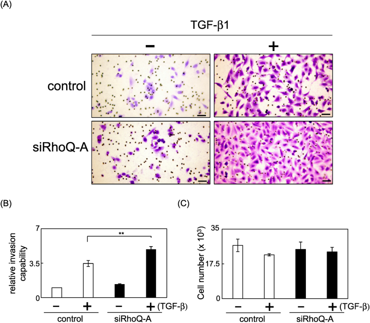
Lung cancer is the leading cause of cancer-related deaths worldwide, and the most common subtype of lung cancer is adenocarcinoma. RhoQ is a Rho family GTPase with primary sequence and structural similarities to Cdc42 and RhoJ. RhoQ is involved in neurite outgrowth via membrane trafficking and is essential for insulin-stimulated glucose uptake in mature adipocytes. However, the function of RhoQ in lung adenocarcinoma (LUAD) remains unclear. In this study, RhoQ siRNAs were introduced into A549 and PC-9 cells. Expression level of EMT-related genes and invasion ability were investigated using Western blot and transwell assay. To examine the relationship between RhoQ expression and prognosis of LUAD, Kaplan-Meier plotter was used. We discovered that suppressing RhoQ expression promoted TGF-β-mediated EMT and invasion in LUAD cell lines. Furthermore, RhoQ knockdown increased Smad3 phosphorylation and Snail expression, indicating that RhoQ was involved in TGF/Smad signaling during the EMT process. Moreover, Kaplan-Meier plotter analysis revealed that low RhoQ levels were associated with poor overall survival in patients with LUAD. In conclusion, these findings shed light on RhoQ's role as a negative regulator of TGF-β-mediated EMT in LUAD.

Keywords: Cdc42, Cell division cycle 42; DMEM, Dulbecco's Modified Eagle's Medium; EMT; EMT, Epithelial-to-Mesenchymal Transition; ERK, Extracellular signal-related kinase; FBS, Fetal Bovine Serum; LUAD, Lung adenocarcinoma; Lung adenocarcinoma; MEK, Mitogen-activated protein kinase kinase; NSCLC, Non-Small-Cell Lung Cancer; RhoQ; SDS, Sodium Dodecyl Sulfate; TGF-β; TGF-β, Transforming Growth Factor-beta; TRITC, tetramethylrhodamine-isothiocyanate; qPCR, Quantitative Polymerase Chain Reaction; rRNA, Ribosomal ribonucleic acid; siRNA, Small Interfering RNA.


Conflict of interest statement

There are no conflicts of interest to declare.

Figures

Fig. 1
Fig. 1
Effect of knockdown of RhoQ on the expression levels of epithelial-to-mesenchymal transition (EMT)-related genes in lung adenocarcinoma cell lines. (A) Cell lysates were prepared from A549 cells transfected with siLuciferase, siRhoQ-A, and siRhoQ-B. Protein levels of RhoQ and β-actin, a loading control, were determined by Western blot analysis. (B) The mRNA levels of RhoQ were assessed by Q-PCR using specific primers for RhoQ. 18S rRNA was used as the internal control for normalization. Each column shows mean ± standard deviation (SD) (n = 3). (C) Cell lysates from control and RhoQ-knockdown cells treated with 1 ng/mL TGF-β1 for 48 h were analyzed on Western blot probed with antibodies against E-cadherin and fibronectin. Each column shows mean ± SD (n = 4). (D) The mRNA levels of E-cadherin and fibronectin were assessed by Q-PCR. Expression of E-cadherin and fibronectin mRNA in control and RhoQ-knockdown cells treated with TGF-β1 for 48 h. Each column shows mean ± SD (n = 3). (E) Cell lysates were prepared from PC-9 cells transfected with siLuciferase and siRhoQ-A. Cell lysates from control and RhoQ-knockdown cells treated with 5 ng/mL TGF-β1 for 48 h were analyzed on Western blot probed with antibodies against E-cadherin and N-cadherin. Each column shows mean ± SD (n = 3). (F) F-actin in RhoQ-knockdown A549 cells treated with 1 ng/mL TGF-β1 for 48 h is visualized with TRITC-conjugated phalloidin. Bars = 25 μm. (G) The ratio of cell major axis and minor axis in A549 cells was measured to evaluate the degree of cell elongation. Each column shows mean ± SD; the number of cells used for these experiments is shown in each bar's parentheses. Significant differences are denoted as **p < 0.01, *p < 0.05.
Fig. 2
Fig. 2
Effect of knockdown of RhoQ on Smad3 phosphorylation and Snail expression during TGF-β-mediated EMT in A549 cells. (A) Samd3 phosphorylation and expression of total Smad3 in control and RhoQ-knockdown cells undergoing TGF-β-mediated EMT were determined by Western blot analysis. Each column shows mean ± SD (n = 4). (B) The expression level of Snail protein in RhoQ-knockdown cells undergoing EMT was determined by Western blot analysis. Each column shows mean ± SD (n = 4). Significant differences are denoted as **p < 0.01.
Fig. 3
Fig. 3
Effect of RhoQ knockdown on the invasion capacity of A549 cells undergoing TGF-β-induced EMT. The cells that invaded the underside of the transwell insert were stained and counted. (A) Representative images of invading cells. Bars = 100 μm. (B) The mean number of invaded cells in the field was calculated. Data were obtained from three independent experiments. Each column shows mean ± SD. (C) Cells undergoing EMT were plated and counted after 24 h to evaluate proliferation capacity. Each column shows mean ± SD (n = 4). Significant differences are denoted as **p < 0.01.
Fig. 4
Fig. 4
Kaplan–Meier analysis in patients with lung adenocarcinoma with high or low expression of RhoQ. Prognosis of patients with lung cancer expression of RhoQ. Correlation between RhoQ expression and prognosis in lung cancers using the online Kaplan–Meier plotter. LUAD is 719 patients (low:361, high:358) and LUSC is 524 patients (low:264, high:260), respectively.

References

    1. Saab S., Zalzale H., Rahal Z., et al. Insights into lung cancer immune-based biology, prevention, and treatment. Front. Immunol. 2020;11:159. - PMC - PubMed
    1. Junyuan L., Yuting Z., Jie T., et al. Heat shock factor 2-binding protein promotes tumor progression via activation of MAPK signaling pathway in lung adenocarcinoma. Bioengineered. 2022;13:10324–10334. - PMC - PubMed
    1. Kundu S.T., Rodriguez B.L., Gibson L.A., et al. The microRNA-183/96/182 cluster inhibits lung cancer progression and metastasis by inducing an interleukin-2-mediated antitumor CD8+ cytotoxic T-cell response. Genes Dev. 2022;36:582–600. - PMC - PubMed
    1. Ramesh V., Brabletz T., Ceppi P. EMT in cancer with repurposed metabolic inhibitors. Trends in Cancer. 2020;6:942–950. - PubMed
    1. Tsubakihara Y., Moustakas A. Epithelial-mesenchymal transition and metastasis under the control of transforming growth factor β. Int. J. Mol. Sci. 2018;19:3672. - PMC - PubMed