. 2023 Mar;75(3):352-363.
doi: 10.1002/art.42353. Epub 2022 Dec 28.

Role of Human Mesenchymal Stem Cells and Derived Extracellular Vesicles in Reducing Sensory Neuron Hyperexcitability and Pain Behaviors in Murine Osteoarthritis

Affiliations

Role of Human Mesenchymal Stem Cells and Derived Extracellular Vesicles in Reducing Sensory Neuron Hyperexcitability and Pain Behaviors in Murine Osteoarthritis

Minji Ai et al. Arthritis Rheumatol. 2023 Mar.

Abstract

Objective: Mesenchymal stem/stromal cells (MSCs) and MSC-derived extracellular vesicles (MSC-EVs) have been reported to alleviate pain in patients with knee osteoarthritis (OA). We undertook this study to determine whether MSCs and/or MSC-EVs reduce OA pain through influencing sensory neuron excitability in OA joints.

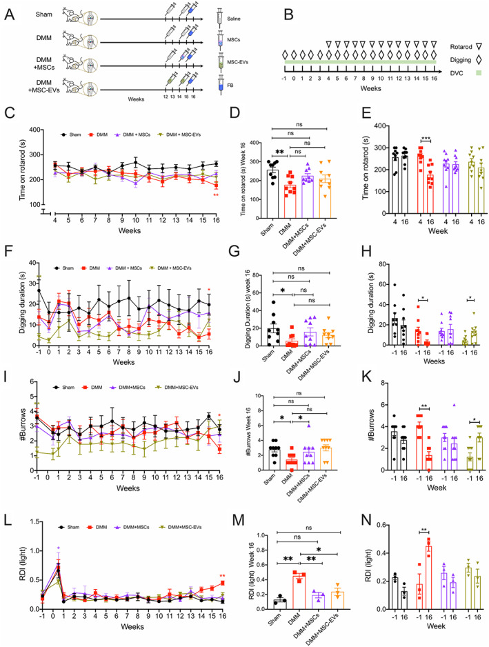
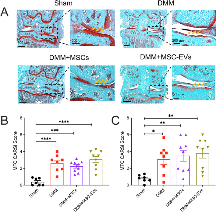
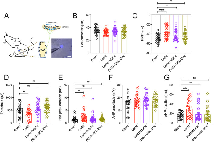
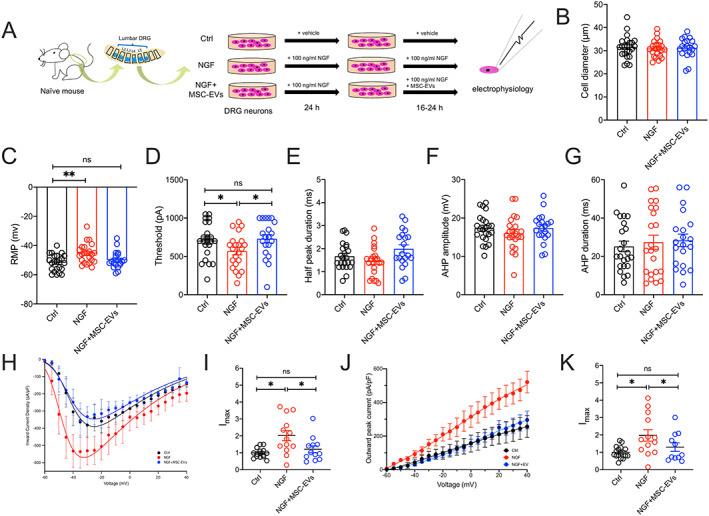
Methods: We induced knee OA in adult male C57BL/6J mice through destabilization of the medial meniscus (DMM) surgery. Mice were sorted into 4 experimental groups with 9 mice per group as follows: unoperated sham, untreated DMM, DMM plus MSC treatment, and DMM plus MSC-EV treatment. Treated mice received either MSCs at week 14 postsurgery or MSC-EVs at weeks 12 and 14 postsurgery. Mouse behavior was evaluated by digging and rotarod tests and the Digital Ventilated Cage system. At week 16, mouse knee joints were harvested for histology, and dorsal root ganglion (DRG) neurons were isolated for electrophysiology. Furthermore, we induced hyperexcitability in DRG neurons in vitro using nerve growth factor (NGF) then treated these neurons with or without MSC-EVs and evaluated neuron excitability.

Results: MSC- and MSC-EV-treated DMM-operated mice did not display pain-related behavior changes (in locomotion, digging, and sleep) that occurred in untreated DMM-operated mice. The absence of pain-related behaviors in MSC- and MSC-EV-treated mice was not the result of reduced joint damage but rather a lack of knee-innervating sensory neuron hyperexcitability that was observed in untreated DMM-operated mice. Furthermore, we found that NGF-induced sensory neuron hyperexcitability is prevented by MSC-EV treatment (P < 0.05 versus untreated NGF-sensitized neurons when comparing action potential threshold).

Conclusion: MSCs and MSC-EVs may reduce pain in OA by direct action on peripheral sensory neurons.


Figures

Figure 1
Figure 1
A, Mice (n = 36) were assigned 1:1:1:1 to sham, destabilization of medial meniscus (DMM) surgery without treatment, DMM with mesenchymal stem/stromal cell (MSC) treatment, or DMM with MSC‐derived extracellular vesicle (MSC‐EV) treatment. Mice in all groups were injected with fast blue (FB) at week 15 postsurgery, and mouse knee joints were harvested at week 16. B, Timeline of behavior tests and time mice spent in Digital Ventilated Cages (DVCs). C–E, Time series comparison of mouse groups from week 4 to week 16 (C), between‐group comparison at week 16 (D), and within‐group comparison between week 4 and week 16 (E) of time mice spent on rotarod. F–N, Time series comparison of mouse groups from presurgery to week 16 (F, I, L), between‐group comparison at week 16 (G, J, M), and within‐group comparison between presurgery and week 16 (H, K, N) of time mice spent digging (F–H), number of burrows mice dug (I–K), and regularity disruption index (RDI) values during lights‐on periods (L–N). Symbols represent individual mice. Dots or bars with whiskers show the mean ± SEM. * = P < 0.05; ** = P < 0.01; *** = P < 0.001 by two‐way analysis of variance (ANOVA) followed by Tukey's post hoc test for time series comparisons, one‐way ANOVA followed by Tukey's post hoc test for between‐group comparisons, or two‐way ANOVA followed by Holm‐Sidak multiple comparisons test for within‐group comparisons. ns = no significant difference. Color figure can be viewed in the online issue, which is available at http://onlinelibrary.wiley.com/doi/10.1002/art.42353/abstract.
Figure 2
Figure 2
Histologic changes in DMM‐operated knee joints at 16 weeks after surgery in mice left untreated or treated with MSCs or MSC‐EVs compared to untreated, unoperated mice in the sham group. Knee joints were assessed for cartilage damage by staining with Safranin O–fast green (A) and Osteoarthritis Research Society International (OARSI) scores of the medial femoral condyle (MFC) (B) and medial tibial condyle (MTC) (C). Yellow arrows indicate intact cartilage in the sham group, whereas in DMM‐operated groups, cartilage loss is evident as reduced red staining. In B and C, symbols represent individual mice; bars with whiskers show the mean ± SEM. * = P < 0.05; ** = P < 0.01; *** = P < 0.001; **** = P < 0.0001 versus sham by one‐way ANOVA followed by Tukey's post hoc test. See Figure 1 for other definitions.
Figure 3
Figure 3
Characteristics of knee‐innervating dorsal root ganglion (DRG) neurons assessed in mice from each group using retrograde fast blue labeling of knee joints (A; bar = 50 μm), with electrophysiology studies comparing diameter (B), resting membrane potential (RMP) (C), threshold of electrical stimulus required for action potential (AP) firing (D), half peak duration (E), afterhyperpolarization (AHP) amplitude (F), and AHP duration (G). Symbols represent individual DRG neurons; bars with whiskers show the mean ± SEM. * = P < 0.05; ** = P < 0.01; *** = P < 0.001 versus sham by one‐way ANOVA followed by Tukey's post hoc test. ns = no significant difference (see Figure 1 for other definitions). Color figure can be viewed in the online issue, which is available at http://onlinelibrary.wiley.com/doi/10.1002/art.42353/abstract.
Figure 4
Figure 4
In vitro studies of DRG neurons performed in cultures with medium alone (control [Ctrl]), 100 ng/ml nerve growth factor β (NGFβ), or NGFβ plus MSC‐EVs (106/ml) for 16–24 hours (h) (A), followed by electrophysiology studies comparing diameter (B), RMP (C), threshold of electrical stimulus required for AP firing (D), half peak duration (E), AHP amplitude (F), and AHP duration (G), as well as peak voltage‐gated inward (H) and outward (J) current density of DRG neurons normalized to cell capacitance in different conditions, and peak voltage‐gated inward (I) and outward (K) current normalized to maximum current density (Imax) of control neurons. Symbols represent individual DRG neuron samples. Bars or horizontal lines with whiskers show the mean ± SEM. * = P < 0.05 and ** = P < 0.01 by one‐way ANOVA followed by Tukey's post hoc test. MSC‐EV = mesenchymal stem/stromal cell–derived extracellular vesicle (see Figure 3 for other definitions). Color figure can be viewed in the online issue, which is available at http://onlinelibrary.wiley.com/doi/10.1002/art.42353/abstract.

References

    1. Safiri S, Kolahi A, Smith E , et al. Global, regional and national burden of osteoarthritis 1990–2017: a systematic analysis of the Global Burden of Disease Study 2017. Ann Rheum Dis 2020;79:819–28. - PubMed
    1. McDonough CM, Jette AM. The contribution of osteoarthritis to functional limitations and disability [review]. Clin Geriatr Med 2010;26:387–99. - PMC - PubMed
    1. Martinez R, Reddy N, Mulligan EP, et al. Sleep quality and nocturnal pain in patients with hip osteoarthritis. Medicine (Baltimore) 2019;98:e17464. - PMC - PubMed
    1. Neogi T. The epidemiology and impact of pain in osteoarthritis. Osteoarthr Cartil 2013;21:1145–53. - PMC - PubMed
    1. Moore N, Pollack C, Butkerait P. Adverse drug reactions and drug‐drug interactions with over‐the‐counter NSAIDs [review]. Ther Clin Risk Manag 2015;11:1061–75. - PMC - PubMed

Publication types

Substances