. 2022 Sep 27;119(39):e2201443119.
doi: 10.1073/pnas.2201443119. Epub 2022 Sep 19.

Scavenger receptor-targeted plaque delivery of microRNA-coated nanoparticles for alleviating atherosclerosis

Affiliations

Scavenger receptor-targeted plaque delivery of microRNA-coated nanoparticles for alleviating atherosclerosis

Qianqian Bai et al. Proc Natl Acad Sci U S A. .

Abstract

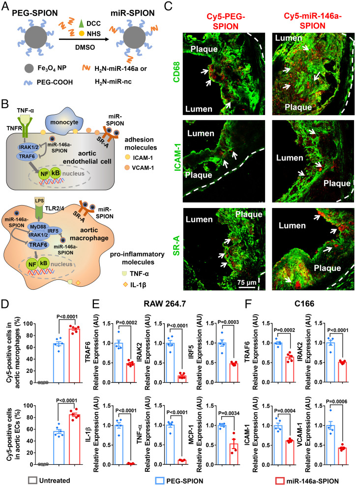
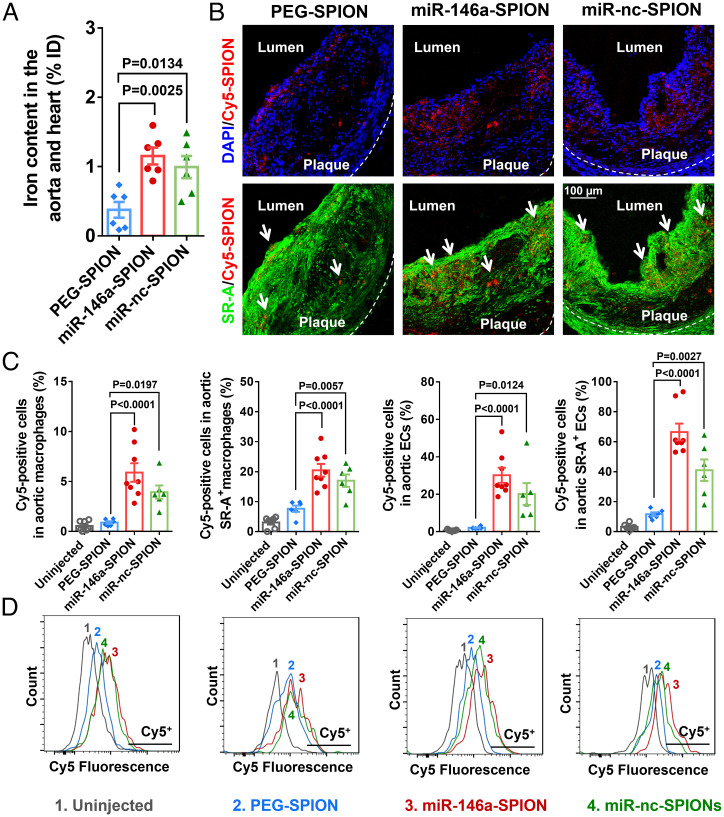
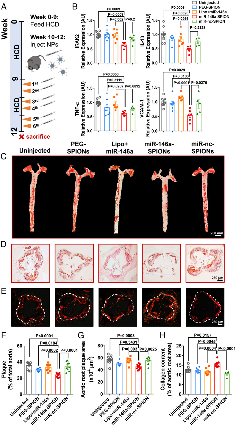
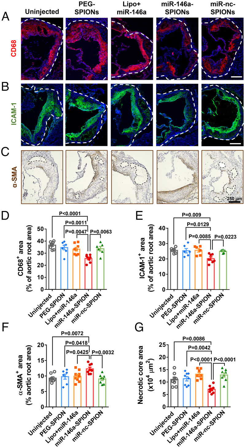
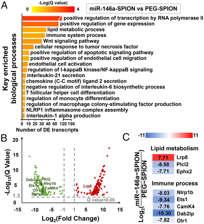
Atherosclerosis treatments by gene regulation are garnering attention, yet delivery of gene cargoes to atherosclerotic plaques remains inefficient. Here, we demonstrate that assembly of therapeutic oligonucleotides into a three-dimensional spherical nucleic acid nanostructure improves their systemic delivery to the plaque and the treatment of atherosclerosis. This noncationic nanoparticle contains a shell of microRNA-146a oligonucleotides, which regulate the NF-κB pathway, for achieving transfection-free cellular entry. Upon an intravenous injection into apolipoprotein E knockout mice fed with a high-cholesterol diet, this nanoparticle naturally targets class A scavenger receptor on plaque macrophages and endothelial cells, contributing to elevated delivery to the plaques (∼1.2% of the injected dose). Repeated injections of the nanoparticle modulate genes related to immune response and vascular inflammation, leading to reduced and stabilized plaques but without inducing severe toxicity. Our nanoparticle offers a safe and effective treatment of atherosclerosis and reveals the promise of nucleic acid nanotechnology for cardiovascular disease.

Keywords: atherosclerosis; cardiovascular disease; gene therapy; nanomedicine; nucleic acid nanotechnology.


Conflict of interest statement

The authors declare no competing interest.

Figures

Fig. 1.
Fig. 1.
Ex vivo binding of miR-146a-SPIONs to endothelial cells and macrophages in atherosclerotic plaque and in vitro down-regulation of genes related to NF-κB pathway by miR-146a-SPIONs in cell lines. (A) Preparation of miR-SPIONs. (B) The NF-κB pathway and miR-146a target in aortic macrophage and EC. (C) Representative confocal images showing the ex vivo association of Cy5-labeled PEG-SPIONs and miR-146a-SPIONs (red; with white arrows) to macrophages, ECs and SR-A in atherosclerotic plaques from aortic root sections. Green pseudo color: CD68 (macrophages), ICAM-1 (ECs) or SR-A. The white dotted lines indicate the border of the vascular wall from aortic roots. (D) Flow cytometric analysis shows the association of Cy5-labeled PEG-SPIONs (blue) and miR-146a-SPIONs (red) to total macrophages and total ECs in the aorta of ApoE−/− mice after 2 h of ex vivo incubation (n = 4–6, across two independent experiments). Statistical significance was calculated by one-way ANOVA with Tukey's test for post hoc analysis. (E and F) In vitro down-regulation of genes in RAW264.7 macrophages and C166 ECs related to the NF-κB proinflammatory pathway 24 h following incubation of miR-146a-SPIONs (red) or PEG-SPION (blue) (n = 5, across two independent experiments). Statistical significance was calculated by Student’s t test. All the data are presented as mean ± SEM.
Fig. 2.
Fig. 2.
Accumulation of miR-146a-SPIONs inside atherosclerotic plaques. (A) ICP-MS measurements of the iron content in the aorta and heart (sites of plaques for ApoE−/− mice) show more abundant accumulation of miR-146a-SPIONs (red) and miR-nc-SPIONs (green) than PEG-SPIONs (blue) 2 h postinjection (n = 6 mice per group, across two independent experiments). (B) Representative confocal images of the aortic root confirm stronger colocalization of Cy5-labeled miR-146a-SPIONs and Cy5-labeled miR-nc-SPIONs than Cy5-PEG-SPIONs (red) with aortic SR-A-rich macrophages or ECs 2 h postinjection. Green pseudo color: SR-A. Blue: DAPI. (C) Flow cytometric analysis of the cells in the aorta reveal stronger association of miR-146a-SPIONs (red) and miR-nc-SPIONs (green) to the aortic cell types than PEG-SPIONs (blue), including total macrophages, SR-A+ macrophages, total ECs, and SR-A+ ECs 2 h postinjection (n = 6–8 mice per group, across two independent experiments). MFI: mean fluorescence intensity. (D) Representative flow cytometry histograms confirm that all types of aortic cells uptake Cy5-labeled miR-146a-SPIONs (red) and Cy5-labeled miR-nc-SPIONs (green) more readily than Cy5-labled PEG-SPIONs (blue) 2 h postinjection. Data are presented as means ± SEM. Statistical significance was calculated by one-way ANOVA with Tukey's test for post hoc analysis.
Fig. 3.
Fig. 3.
miR-146a-SPIONs reduced and stabilized atherosclerotic plaques and inhibited genes linked to atherogenesis. (A) After 9 wk of HCD, the ApoE−/− mice received two weekly i.v. injections from weeks 10–12 while continuing the HCD. (B) qRT-PCR analysis of aortic RNA reveals significant down-regulation of genes targeted by miR-146a (IRAK2) and genes downstream of the NF-κB pathway (IL-1β, TNF-α, and VCAM-1) by miR-146a-SPIONs. For the miR-146a-SPION group, representative images of (C) Oil Red O (ORO)-stained intact aortas and (D) ORO-stained sections of aortic roots indicate most substantial reduction in the plaque fat content (red), and (E) Sirius Red-stained aortic roots reveal most abundant accumulation of collagen (red). For the miR-146a-SPION group, the (F) fractional plaque area in total aorta (n = 8–10 mice per group, across three independent experiments) is the lowest, (G) plaque area in the aortic root is the lowest, and (H) collagen content in aortic root is the highest. Data of aortic root sections were chosen from n = 6 images per mouse from n = 6–8 mice per group. Data are presented as means ± SEM. Statistical significance was calculated by one-way ANOVA with Tukey's test for post hoc analysis.
Fig. 4.
Fig. 4.
miR-146a-SPIONs inhibited the morphological markers of atherosclerotic plaques. Representative confocal images of the sections of aortic roots reveal most apparent reduction in (A) CD68 (marker of monocytes and macrophages; red pseudo color) for the miR-146a-SPION group and (B) ICAM-1 (marker of ECs; green pseudo color). Blue: DAPI. (C) Representative IHC images of the sections of aortic roots reveal most apparent enhancement in of α-SMA (highly expressed by smooth muscle cells; brown) for the miR-146a-SPION group. The dotted region indicates the necrotic core of the plaque. For the miR-146a-SPION group, (D) CD68-positive area in the aortic root is the lowest, (E) ICAM-1-positive area in the aortic root is the lowest, (F) α-SMA-positive area in the aortic root is the highest, and (G) area of necrotic core in the aortic root is the lowest. Data of aortic root sections were from an average of six images per mouse, from n = 6–8 mice per group (across three independent experiments). Data are presented as means ± SEM. Statistical significance was calculated by one-way ANOVA with Tukey's test for post hoc analysis.
Fig. 5.
Fig. 5.
miR-146a-SPIONs modulated the expression of genes related to immune response pathways and vascular inflammation in comparison to PEG-SPIONs. (A) Key enriched biological processes found by comparing the “miR-146a-SPION” group (n = 3) to the “PEG-SPION” group (n = 3). (B) Volcano plot showing the distribution of DETs by comparing the “miR-146a-SPION” group to the “PEG-SPION” group (absolute fold change > 2). (C) Representative DETs found in the comparison between “miR-146a-SPION” and “PEG-SPION” groups indicate the effect of miR-146a-SPION treatment on gene expression.

References

    1. Andreou I., Sun X., Stone P. H., Edelman E. R., Feinberg M. W., miRNAs in atherosclerotic plaque initiation, progression, and rupture. Trends Mol. Med. 21, 307–318 (2015). - PMC - PubMed
    1. Hansson G. K., Libby P., The immune response in atherosclerosis: A double-edged sword. Nat. Rev. Immunol. 6, 508–519 (2006). - PubMed
    1. Liu M. W., et al. , Trapidil in preventing restenosis after balloon angioplasty in the atherosclerotic rabbit. Circulation 81, 1089–1093 (1990). - PubMed
    1. Sarembock I. J., et al. , Influence of inflation pressure and balloon size on the development of intimal hyperplasia after balloon angioplasty. A study in the atherosclerotic rabbit. Circulation 80, 1029–1040 (1989). - PubMed
    1. Fleg J. L., et al. , Effect of statins alone versus statins plus ezetimibe on carotid atherosclerosis in type 2 diabetes: The SANDS (Stop Atherosclerosis in Native Diabetics Study) trial. J. Am. Coll. Cardiol. 52, 2198–2205 (2008). - PMC - PubMed

Publication types