. 2022 Nov 1;34(12):2275-2296.
doi: 10.1162/jocn_a_01915.

The Angular Gyrus as a Hub for Modulation of Language-related Cortex by Distinct Prefrontal Executive Control Regions

Affiliations

The Angular Gyrus as a Hub for Modulation of Language-related Cortex by Distinct Prefrontal Executive Control Regions

Hyojeong Kim et al. J Cogn Neurosci. .

Abstract

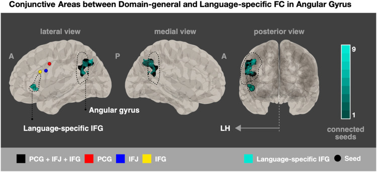
It has become clear in recent years that reading, while relying on domain-specific language processing regions, also involves regions that implement executive processes more broadly. Such executive control is generally considered to be implemented by prefrontal regions, which exert control via connectivity that allows them to modulate processing in target brain regions. The present study examined whether three previously identified and distinct executive control regions in the pFC [Wang, K., Banich, M. T., Reineberg, A. E., Leopold, D. R., Willcutt, E. G., Cutting, L. E., et al. Left posterior prefrontal regions support domain-general executive processes needed for both reading and math. Journal of Neuropsychology, 14, 467-495, 2020] show similar patterns of functional connectivity (FC) during a reading comprehension task as compared with a symbol identification condition. Our FC results in a sample of adolescents (n = 120) suggest all three regions commonly show associations with activity in "classic" left hemisphere reading areas, including the angular and supramarginal gyri, yet each exhibits differential connectivity as well. In particular, precentral regions show differential FC to parietal portions of the dorsal language stream, the inferior frontal junction shows differential FC to middle temporal regions of the right hemisphere and other regions involved in semantic processing, and portions of the inferior frontal gyrus show differential FC to an extensive set of right hemisphere prefrontal regions. These results suggest that prefrontal control over language-related regions occurs in a coordinated yet discrete manner.


Figures

<b>Figure 1.</b>
Figure 1.
Two models of prefrontal connectivity to posterior brain regions involved in language processing. (A) A language-based model posits distinct and nonoverlapping dorsal and ventral pathways (i.e., white-matter fiber tracts) connecting prefrontal and posterior regions of cortex, mainly in the temporal lobe, that are involved in language processing (based on Friederici, 2015). (B) A domain-general model proposes that angular gyrus acts as a central hub of connectivity for a variety of prefrontal regions but then branching connectivity from the angular gyrus is specific for distinct prefrontal regions (Seghier, 2013). The numbers on the cortex represent Brodmann areas that are separated by dashed lines. SFG/MFG/IFG = superior/middle/inferior frontal gyrus; PC = parietal cortex; MTG/ITG = middle/inferior temporal gyrus. The brain images were created using the CONN toolbox.
<b>Figure 2.</b>
Figure 2.
Psychological factor and physiological factor for gPPI. (A) Experimental condition and design. There are four conditions, which are passages, words, symbols, and pictures, in the reading comprehension task across three runs with a block design. The psychological factor for the gPPI model is shown on the bottom. (B) Seeds. The three domain-general cognitive control seeds were extracted from the three-way conjunction analysis for the physiological factor: left PCG (in red [−46, 2, 38], BA 6), IFJ (in blue [−46, 8, 26], BA 9), and IFG (in yellow [−48, 18, 24], BA 9/44). These seeds were not anatomically overlapping. In addition, the language-identified IFG-seeds were created from the language network in CONN with a searchlight method, and the seeds that had functional connections to angular gyrus are presented.
<b>Figure 3.</b>
Figure 3.
FC results across PCG, IFJ, and IFG seeds. Conjunctive maps of passages versus symbols contrast for greater FC for passages than symbols (A) and greater FC for symbols than passages (B) are shown with assigned colors detailed in the color diagram at the bottom. Three primary colors (red, blue, and yellow) indicate the unique FC areas from each seed, and combined colors indicate the conjunction areas for either two seeds (2way; purple, orange, green) or three seeds (3way; black). The three seeds are presented in circles with their defined colors (LH, lateral) on the volumetric brain. (C–E) The FC results of each seed are shown for greater FC for passages than symbols (in pink) and greater FC for symbols than passages (in sky blue): (C) PCG; (D) IFJ; and (E) IFG. The brain images were created using the CONN toolbox.
<b>Figure 4.</b>
Figure 4.
Different FC to the angular gyrus for executive than language-identified prefrontal seeds. The greater FC areas of the language-identified seeds for the passages as compared with symbol conditions found in angular gyrus are displayed in the left hemisphere (in teal color). The gradation of colors indicates the number of seeds that were functionally connected to the areas in angular gyrus (e.g., the brightest teal represented regions connected to nine of the language-identified left IFG seeds). The 10 seeds out of a total 69 seeds examined that had FC to a portion of the left angular gyrus are presented (also in teal) within the language-identified LIFG ROI (in dashed line; defined in the CONN network). The domain-general seeds are also presented in circles with their defined colors for the 3way-conjunction area in the angular gyrus (in black). The left angular gyrus defined in the FMRIB Software Library Harvard–Oxford Atlas is also outlined by the black dashed line.

References

    1. Aron, A. R., Robbins, T. W., & Poldrack, R. A. (2004). Inhibition and the right inferior frontal cortex. Trends in Cognitive Sciences, 8, 170–177. 10.1016/j.tics.2004.02.010, - DOI - PubMed
    1. Aron, A. R., Robbins, T. W., & Poldrack, R. A. (2014). Inhibition and the right inferior frontal cortex: One decade on. Trends in Cognitive Sciences, 18, 177–185. 10.1016/j.tics.2013.12.003, - DOI - PubMed
    1. Binder, J. R., Desai, R. H., Graves, W. W., & Conant, L. L. (2009). Where is the semantic system? A critical review and meta-analysis of 120 functional neuroimaging studies. Cerebral Cortex, 19, 2767–2796. 10.1093/cercor/bhp055, - DOI - PMC - PubMed
    1. Braga, R. M., DiNicola, L. M., Becker, H. C., & Buckner, R. L. (2020). Situating the left-lateralized language network in the broader organization of multiple specialized large-scale distributed networks. Journal of Neurophysiology, 124, 1415–1448. 10.1152/JN.00753.2019, - DOI - PMC - PubMed
    1. Calhoun, V. D., Wager, T. D., Krishnan, A., Rosch, K. S., Seymour, K. E., Nebel, M. B., et al. (2017). The impact of T1 versus EPI spatial normalization templates for fMRI data analyses. Human Brain Mapping, 38, 5331–5342. 10.1002/hbm.23737, - DOI - PMC - PubMed

Publication types