. 2023 Jan 12;141(2):135-146.
doi: 10.1182/blood.2022016504.

Missense mutations in PIEZO1, which encodes the Piezo1 mechanosensor protein, define Er red blood cell antigens

Affiliations

Missense mutations in PIEZO1, which encodes the Piezo1 mechanosensor protein, define Er red blood cell antigens

Vanja Karamatic Crew et al. Blood. .

Abstract

Despite the identification of the high-incidence red cell antigen Era nearly 40 years ago, the molecular background of this antigen, together with the other 2 members of the Er blood group collection, has yet to be elucidated. Whole exome and Sanger sequencing of individuals with serologically defined Er alloantibodies identified several missense mutations within the PIEZO1 gene, encoding amino acid substitutions within the extracellular domain of the Piezo1 mechanosensor ion channel. Confirmation of Piezo1 as the carrier molecule for the Er blood group antigens was demonstrated using immunoprecipitation, CRISPR/Cas9-mediated gene knockout, and expression studies in an erythroblast cell line. We report the molecular bases of 5 Er blood group antigens: the recognized Era, Erb, and Er3 antigens and 2 novel high-incidence Er antigens, described here as Er4 and Er5, establishing a new blood group system. Anti-Er4 and anti-Er5 are implicated in severe hemolytic disease of the fetus and newborn. Demonstration of Piezo1, present at just a few hundred copies on the surface of the red blood cell, as the site of a new blood group system highlights the potential antigenicity of even low-abundance membrane proteins and contributes to our understanding of the in vivo characteristics of this important and widely studied protein in transfusion biology and beyond.


Conflict of interest statement

Conflict-of-interest disclosure: The authors declare no competing financial interests.

Figures

None
Graphical abstract
Figure 1.
Figure 1.
PIEZO1 gene and Piezo1 protein cartoons. (A) Scale representation of PIEZO1 gene (NM_001142864.4), highlighting exons 45 to 50 (shown in blue), which encode the large extracellular loop (amino acids 2198-2431) of the Piezo1 protein. (B) Piezo1 protein (Uniprot Q92508) cartoon generated in Protter with large extracellular loop (amino acids 2198-2431) shown in blue.
Figure 2.
Figure 2.
Mutations in PIEZO1 gene (exons 45-50) encoding amino acid substitutions in Piezo1 protein extracellular loop. (A) PIEZO1 genotypes observed in Er(a−b+) and Er(a−b−) individuals. PIEZO1 exons 45 to 50 are shown (regions coding for extracellular domain shown in blue). Mutations are highlighted in pink (7180G>A) and yellow (7174G>A), seen in homozygous, heterozygous, and compound heterozygous states in individuals tested as shown. Individual P10 has nonsense mutation in exon 38 (not shown) predicted to encode truncated protein (exons colored gray). (B) PIEZO1 alleles observed in Er variant individuals. PIEZO1 exons 45 to 50 are shown. Mutations are highlighted in purple (7219G>C), green (7219G>A), and brown (6734G>A). All mutations were homozygous in the indicated individuals. (C) All mutations shown in panels A and B encode amino acid substitutions in the extracellular domain of Piezo1 protein (amino acids 2198-2431) as shown (colors as in 2A and 2B).
Figure 3.
Figure 3.
Anti-Er alloantibodies are specific for antigenic sites on Piezo1. Flow cytometry histograms illustrate cell surface labeling of indicated wild-type or mutant Piezo1 constructs overexpressed in an endogenous Piezo1 knockout (KO) BEL-A cell line using antibodies eluted from plasma of individuals (P3, row A; P11, row B; P12, row C; and P13, row D) with mutations in Piezo1 as labeled. BEL-A Piezo1 KO cells and endogenous Piezo1 are shown in column 1, with overexpressed wild-type Piezo1 in column 2. Columns 3 to 7 show overexpressed mutant Piezo1 constructs as labeled. Results are summarized in the grid below histograms. O-E WT, overexpressed wild-type; +, positive; −, negative; (+), weakly positive.
Figure 4.
Figure 4.
Evaluation of Piezo1 activity in BEL-A overexpressed wild-type and mutant cells using patch clamp. Raw traces of current amplitudes (A) and time course of the experiment (B) at 80 and −100 mV from an example overexpressed wild-type (WT) Piezo1 cell obtained before (precompound, red trace/symbols) and after (compound, blue trace/symbols) addition of 10 μM Yoda1, using a voltage ramp protocol (−100 to 80 mV, 400 ms, holding potential −30 mV) and inhibited by 30 μM GdCl3 (block, green trace/symbols). (C) Evaluation of current increase on Yoda1 addition (Yoda1 induced current, IYoda1− IES, pA/pF) of responding/nonresponding cells identified from BEL-A overexpressed wild-type and mutant Piezo1 constructs (n = 11/77 overexpressed WT; n = 21/84 Glu2392Lys; n = 13/80 Gly2394Ser; n = 15/89 Glu2407Gln; n = 15/80 Glu2407Lys; n = 19/85 Arg2245Gln). (D) Evaluation of current increase on Yoda1 addition in the presence of TRAM-34 (Yoda1 + TRAM-34 induced current, IYoda1− IES, pA/pF) of responding cells identified from BEL-A overexpressed wild-type and mutant Piezo1 constructs (n = 48/82 overexpressed WT; n = 54/86 Glu2392Lys; n = 62/87 Gly2394Ser; n = 47/93 Glu2407Gln; n = 47/77 Glu2407Lys; n = 45/84 Arg2245Gln).
Figure 5.
Figure 5.
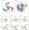
Modeling of Piezo1 protein and location of Er antigenic sites. A homology model for the human Piezo1 sequence was generated using coordinates form PDB ID 5Z10 and inserted into a model membrane for subsequent molecular dynamics calculations for a total trajectory time of 100 ns. (A) Top view on extracellular side of equilibrated Piezo1 trimer (composed of 3 chains: red, green, and blue). Lipids have been omitted from the illustration for simplicity. The residues encompassing the antigenic sites are shown as spheres (colored by atom type) located in and around the center of the trimer. (B) Side-view slice of the Piezo1 trimer with the transmembrane section of 1 monomer (red) removed to allow view on center. Representation is the same as in panel A. Side chains of antigenic sites (Era/Erb, Er4, and Er5) are highlighted for the red monomer. (C-E) Era/Erb loops as predicted by molecular dynamics calculations. The backbones of loops encompassing residues 2384 to 2408 are shown in green with the remainder of the protein trimer in beige. The conformation (in green) is representative of the highest populated cluster in the trajectory. The first row represents a side view onto the central extracellular domain, and below it is a depiction of the apical view on the membrane plane as indicated in figure panels. (C) Era representative loop. (D) Erb representative loop. (E) Glu2392Lys representative loop.

Comment in

References

    1. International Society of Blood Transfusion (ISBT) Red cell immunogenetics and blood group terminology. https://www.isbtweb.org/working-parties/red-cell-immunogenetics-and-bloo...
    1. Daniels GL, Judd WJ, Moore BP, et al. A 'new' high frequency antigen Era. Transfusion. 1982;22(3):189–193. - PubMed
    1. Hamilton JR, Beattie KM, Walker RH, Hartrick MB. Erb, an allele to Era, and evidence for a third allele, Er. Transfusion. 1988;28(3):268–271. - PubMed
    1. Arriaga F, Mueller A, Rodberg K, et al. A new antigen of the Er collection. Vox Sang. 2003;84(2):137–139. - PubMed
    1. Coste B, Mathur J, Schmidt M, et al. Piezo1 and Piezo2 are essential components of distinct mechanically activated cation channels. Science. 2010;330(6000):55–60. - PMC - PubMed

Publication types