Review
. 2022 Oct:84:104260.
doi: 10.1016/j.ebiom.2022.104260. Epub 2022 Sep 19.

Therapeutic strategies in ischemic cardiomyopathy: Focus on mitochondrial quality surveillance

Affiliations
Review

Therapeutic strategies in ischemic cardiomyopathy: Focus on mitochondrial quality surveillance

Xing Chang et al. EBioMedicine. 2022 Oct.

Abstract

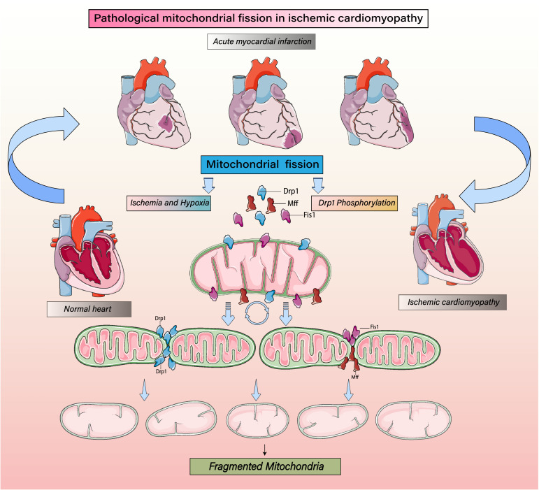
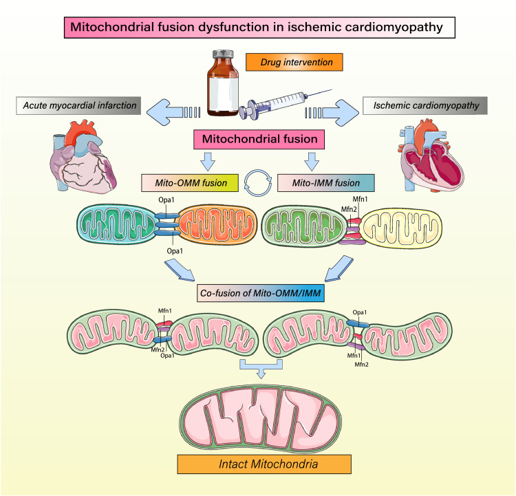
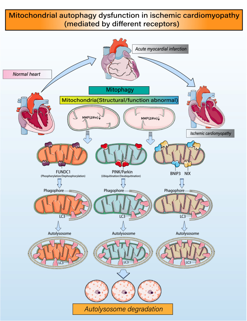
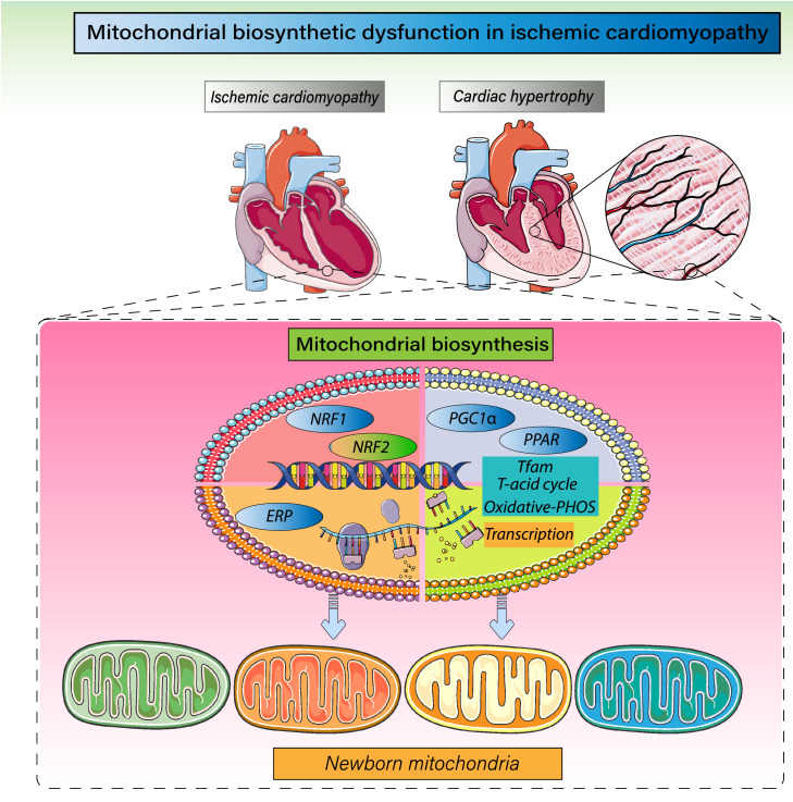
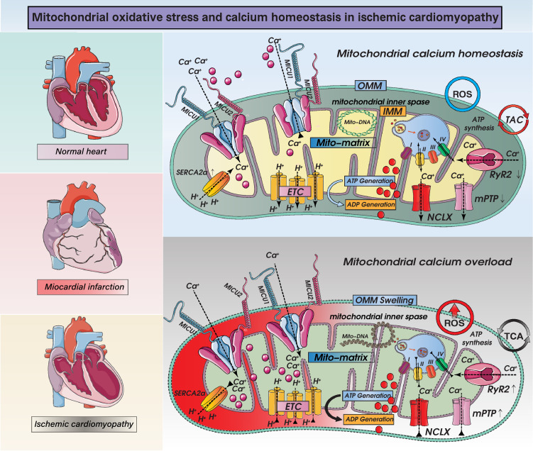
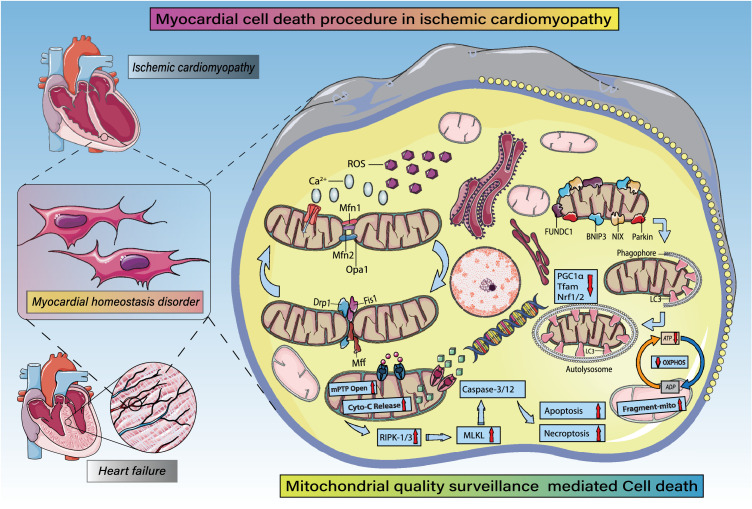
Despite considerable efforts to prevent and treat ischemic cardiomyopathy (ICM), effective therapies remain lacking, in part owing to the complexity of the underlying molecular mechanisms, which are not completely understood yet. It is now widely thought that mitochondria serve as "sentinel" organelles that are capable of detecting cellular injury and integrating multiple stress signals. These pathophysiological activities are temporally and spatially governed by the mitochondrial quality surveillance (MQS) system, involving mitochondrial dynamics, mitophagy, and biogenesis. Dysregulation of MQS is an early and critical process contributing to mitochondrial bioenergetic dysfunction and sublethal injury to cardiomyocytes during ICM. An improved understanding of the pathogenesis of ICM may enable the development of novel preventive and therapeutic strategies aimed at overcoming the challenge of myocardial ischemia and its cardiovascular sequelae. This review describes recent research on the protective effects of MQS in ICM and highlights promising therapeutic targets.

Keywords: Cardiomyocyte; Ischemic cardiomyopathy; Mitochondrial biogenesis; Mitochondrial dynamics; Mitochondrial quality surveillance; Mitophagy.


Conflict of interest statement

Declaration of interests The authors have no conflicts of interest to declare.

Figures

Figure 1
Figure 1
Regulatory mechanism of mitochondrial fission on mitochondrial homeostasis. Mitochondrial fission induced by ICM. Ischemic/hypoxic stress increases phosphorylation of the mitochondrial fission protein Drp1 as well as the expression levels of the Drp1 receptor proteins fission protein 1(Fis1) and mitochondrial fission factor (Mff). Together these proteins mediate typical mitochondrial fission. Mitochondrial fission increases a precursor to mitophagy when cells are subjected to various stresses. This is related to the accumulation of various markers of mitochondrial autophagy. Drp1 phosphorylation induces mitochondrial fission by mediating its oligomerization around the mitochondrial outer membrane after recruitment and stabilization of Fis1 and Mff. Increasing levels of phosphorylated Drp1 increase mitochondrial fission, leading to their fragmentation, which will reduce both energy metabolism and cellular activity and stimulate mitophagy.
Figure 2
Figure 2
Regulation of mitochondrial fusion and mitochondrial homeostasis. Mitochondrial fusion is highly important for maintaining mitochondrial homeostasis and the integrity and quality of mitochondrial structure to ensure that proper energy metabolism is maintained even during ischemia/hypoxia. Mitochondrial fusion is accomplished through the coordinated execution of separate mechanisms underlying fusion of the mitochondrial inner and outer membranes. Mitochondrial inner membrane fusion is mediated mainly by optic atrophy 1 (OPA1) protein, while outer membrane fusion is mediated mainly by mitofusin 1 (Mfn1) and Mfn2.
Figure 3
Figure 3
Regulatory mechanisms governing mitophagy and mitochondrial homeostasis. Under ischemia/hypoxic conditions, fragmented mitochondria or mitochondria with structural damage caused by mitochondrial fission will experience a loss of mitochondrial membrane potential (ΔΨm). Mitochondria with low membrane potentials recruit FUN14 domain-containing 1(FUNDC1), which phosphorylates/dephosphorylates various proteins, leading to high expression of LC3 and formation of mitochondrial autophagic lysosomes. Phosphatase and tensin homolog-induced putative kinase 1 (PINK1) is also translocated to mitochondria, where it induces Parkin to ubiquitinate proteins in the mitochondrial outer membrane. The ubiquitinated mitochondria interact with LC3 on lysosomes to form autophagosomes. In addition, BCL-2/adenovirus E1B 19 kDa protein-interacting protein 3 (BNIP3) and NIX contain an LC3 interaction motif (LIR), which directly binds LC3 and promotes formation of autophagic lysosomes. Through these processes, the mitochondrial self-renewal and homeostasis necessary for cardiomyocyte activity are achieved.
Figure 4
Figure 4
Regulatory mechanism governing mitochondrial biogenesis and mitochondrial homeostasis. Mitochondrial biosynthesis mainly occurs in the late stage of mitochondrial fission and mitophagy. PGC1α is the primary regulatory factor controlling mitochondrial biogenesis and is situated upstream of the mitochondrial biosynthesis reticular regulatory system. It is the hub connecting external signals to internal functional regulation within mitochondria. PGC1α promotes transcription of mitochondrial transcription factor A (TFAM) by activating nuclear respiratory factors 1 and 2 (Nrf-1 and Nrf-2) and regulates the citric acid and tricarboxylic acid cycles and, in turn, oxidative phosphorylation. In addition, ERP and PPAR-γ are members of the nuclear receptor transcription factor superfamily that act to regulate expression of target genes and the levels of TFAM transcription and mitochondrial biosynthesis.
Figure 5
Figure 5
Mechanisms of mitochondrial calcium homeostasis and ROS-mediated mitochondrial oxidative stress injury and mitochondrial dysfunction in ICM. Impact of ROS overproduction-activated oxidative stress injury in ICM on mitochondrial homeostasis. Intrinsic and extrinsic stress signals, such as hypoxia, ischemia, reperfusion, and inflammation, can mediate overproduction of ROS and can lead to mitochondrial oxidative stress damage. When mitochondria are damaged excessively, mitochondrial outer membrane permeabilization (OMM) is dysfunctional, and mitochondrial permeability transition pore (mPTP) is abnormally opened. On the other hand, mPTP opening induces necrosis or necroptosis, accompanied by oxidative phosphorylation dysfunction and dysregulation of the tricarboxylic acid cycle. A range of dysfunctions also induces abnormal calcium signaling, excess ROS production and mitochondrial respiratory chain dysfunction and related protein gene expression changes. Disruption of the mitochondrial calcium cycle has important linkages with mitochondrial uniporter complexes (consisting of MCU, EMRE, MICU1, MICU2, MICU3, MCUB and MCUR1), NCLX, LETM1. And calcium transport in mitochondria is also closely related to ryanodine receptors and mitochondrial permeability transition pore. The normal operation of the above mechanisms(mitochondrial calcium homeostasis and oxidative stress injury) directly or indirectly affects the normal operation of mitochondrial respiratory chain function and mitochondrial energy metabolism function.
Figure 6
Figure 6
MQS in cardiomyocyte fate regulation (apoptosis, necrosis, necroptosis, pyroptosis, autophagic cell death). MQS fully participates in apoptotic or necrotic cardiomyocyte death. Under ischemic/hypoxic stress, intracellular ROS are overproduced and accompanied by intracellular Ca2+ overload. Ischemic stress also breaks the balance between Drp1/FIS1/Mff-mediated mitochondrial fission and OPA1/Mfn1/Mfn2-mediated mitochondrial fusion, which leads to mitochondrial fragmentation and structurally abnormal mitochondria. Mitochondrial autophagy mediated by FUNDC1/Parkin /BNIP3 and NIX is enhanced, leading to PGC1α- and Nrf1/2-mediated decreases in mitochondrial biosynthesis, abnormal opening of mPTPs, and excessive release of Cyto-c into the cytosol. Dysregulation of the mitochondrial electron transport chain and tricarboxylic acid cycle leads to insufficient ATP production. Expression levels of RIPK3 and MLKL are increased, as is activation of caspase-3/-12 and levels of cardiomyocyte.

References

    1. Hearse DJ. Myocardial ischaemia: can we agree on a definition for the 21st century? Cardiovasc Res. 1994;28(12):1737–1744. discussion 45-6. - PubMed
    1. Cabac-Pogorevici I, Muk B, Rustamova Y, et al. Ischaemic cardiomyopathy. Pathophysiological insights, diagnostic management and the roles of revascularisation and device treatment. Gaps and dilemmas in the era of advanced technology. Eur J Heart Fail. 2020;22(5):789–799. - PubMed
    1. Kim H, Scimia MC, Wilkinson D, et al. Fine-tuning of Drp1/Fis1 availability by AKAP121/Siah2 regulates mitochondrial adaptation to hypoxia. Mol Cell. 2011;44(4):532–544. - PMC - PubMed
    1. Rabinovich-Nikitin I, Rasouli M, Reitz CJ, et al. Mitochondrial autophagy and cell survival is regulated by the circadian clock gene in cardiac myocytes during ischemic stress. Autophagy. 2021;17(11):3794–3812. - PMC - PubMed
    1. Ahuja P, Zhao P, Angelis E, et al. Myc controls transcriptional regulation of cardiac metabolism and mitochondrial biogenesis in response to pathological stress in mice. J Clin Invest. 2010;120(5):1494–1505. - PMC - PubMed