. 2023 Jan;50(1):121-130.
doi: 10.1111/jcpe.13729. Epub 2022 Oct 12.

Tobacco smoke exacerbates Filifactor alocis pathogenicity

Affiliations

Tobacco smoke exacerbates Filifactor alocis pathogenicity

Mina M Z Iskander et al. J Clin Periodontol. 2023 Jan.

Abstract

Aim: Filifactor alocis has recently emerged as a periodontal pathobiont that appears to thrive in the oral cavity of smokers. We hypothesized that identification of smoke-responsive F. alocis genes would provide insight into adaptive strategies and that cigarette smoke would enhance F. alocis pathogenesis in vivo.

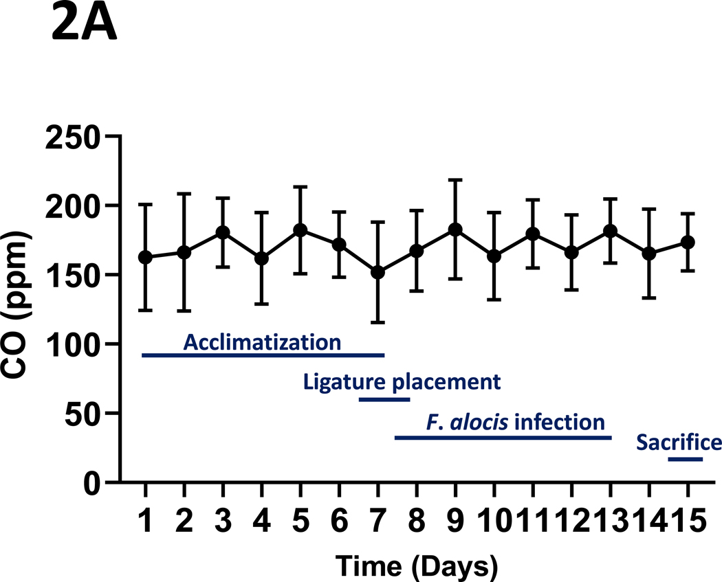
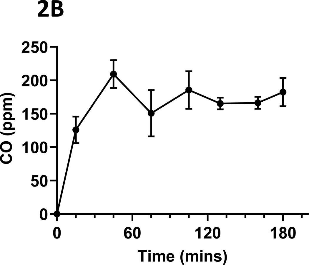
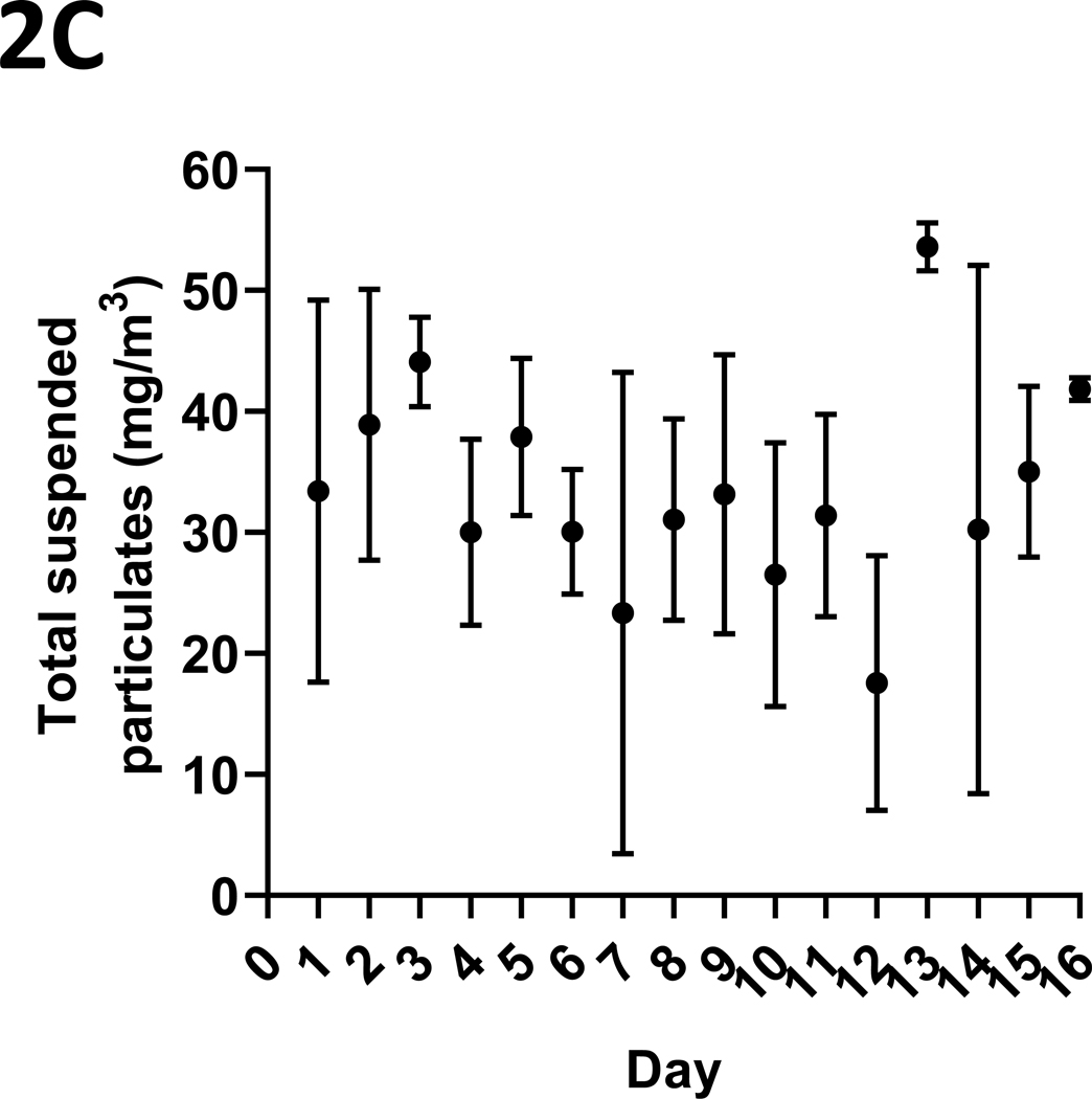
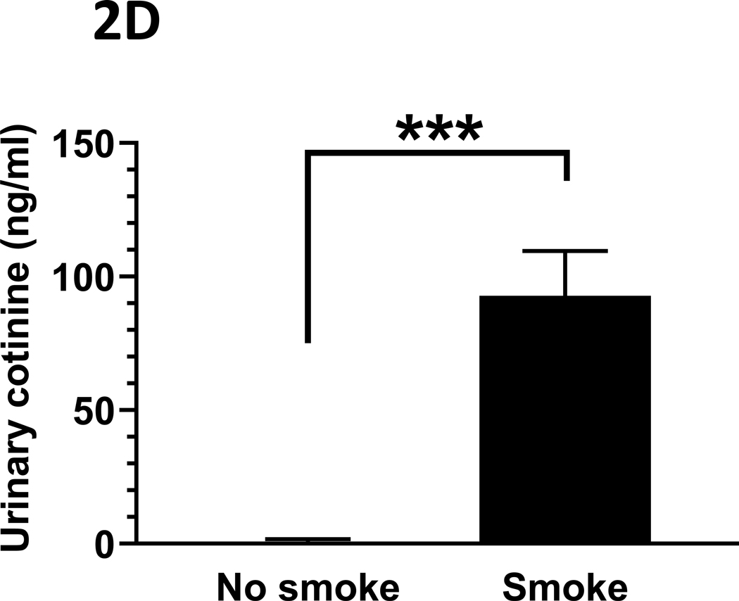
Materials and methods: F. alocis was grown in vitro and cigarette smoke extract-responsive genes determined by RNAseq. Mice were exposed, or not, to mainstream 1R6F research cigarette smoke and infected with F. alocis, or not, in an acute ligature model of periodontitis. Key clinical, infectious, and immune data were collected.

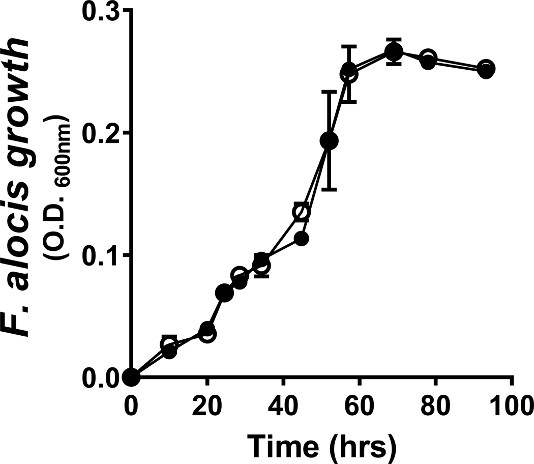
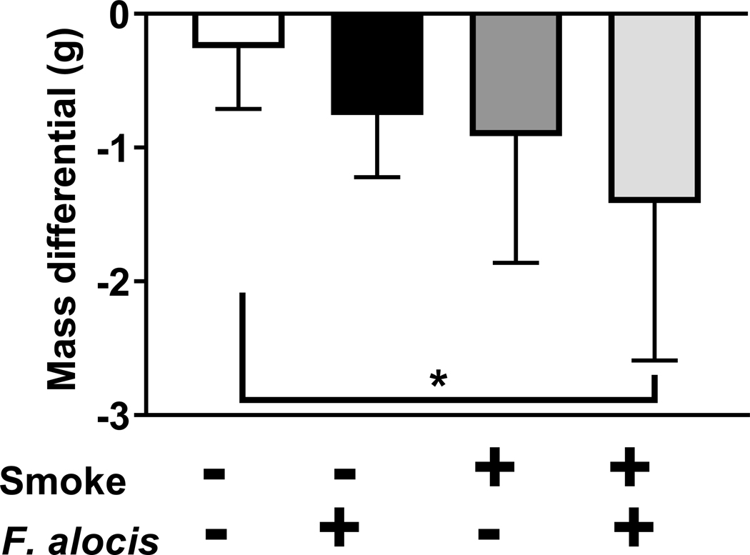
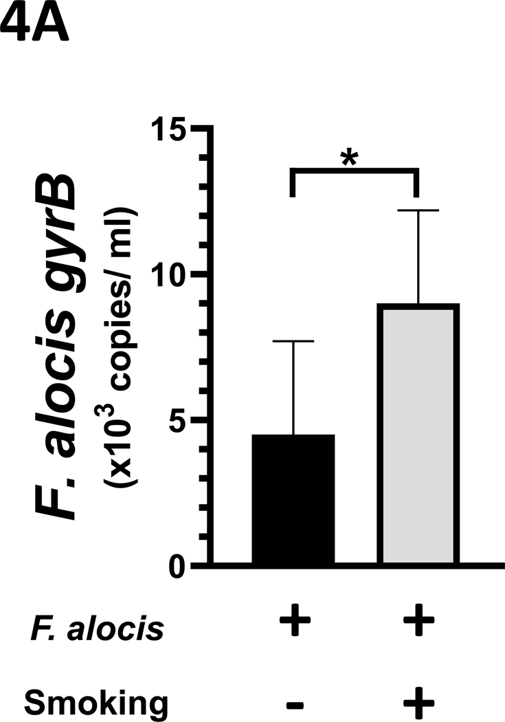
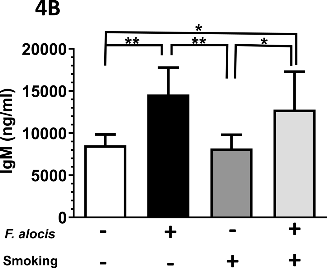
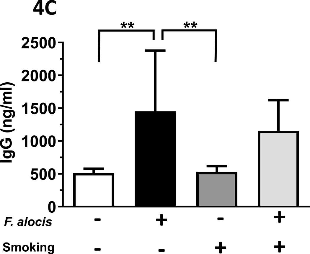
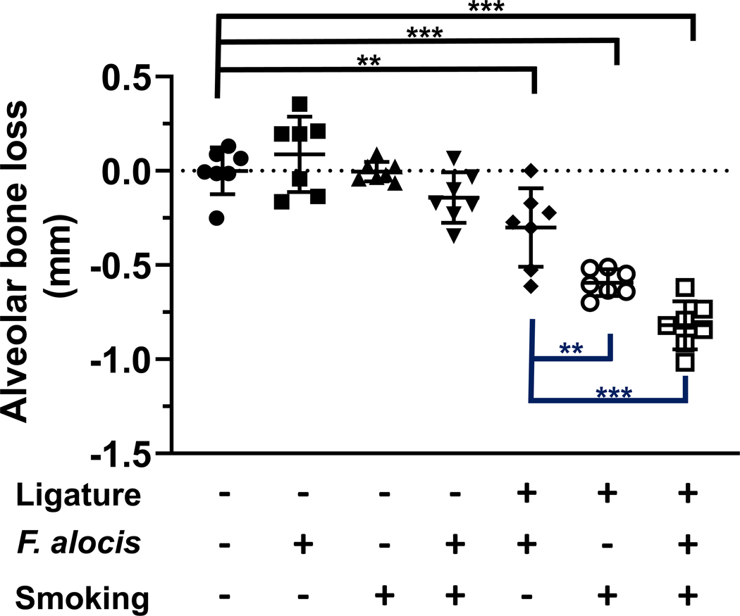
Results: In culture, F. alocis growth was unaffected by smoke conditioning and only a small number of genes were specifically regulated by smoke exposure. Reduced murine mass, differences in F. alocis-cognizant antibody production, and altered immune profiles as well as altered alveolar bone loss were all attributable to smoke exposure and/or F. alocis infection in vivo.

Conclusions: F. alocis is well-adapted to tobacco-rich conditions and its pathogenesis is enhanced by tobacco smoke exposure. A smoke-exposed ligature model of periodontitis shows promise as a tool with which to further unravel mechanisms underlying tobacco-enhanced, bacteria-induced disease.

Keywords: Filifactor alocis; alveolar bone loss; experimental periodontitis; microbiology; tobacco smoking.


Conflict of interest statement

Conflict of Interest statement: The authors have no conflicts of interest to declare.

Figures

Figure 1:
Figure 1:. Cigarette smoke extract does not influence the growth of F. alocis in culture
There were no significant differences in the growth characteristics of F. alocis cultured anaerobically in BHI (black circles) or BHI conditioned with 1R6F cigarette smoke (1000 ng/ml nicotine equivalents) (white circles), as assessed spectrophotometrically (O.D.600nm). Data represent mean (s.d.) values.
Figure 2:
Figure 2:. Tobacco smoke exposure indices.
Mice were exposed to mainstream cigarette smoke generated from 1R6F research cigarettes using a Teague TE-10C cigarette smoke inhalation exposure system for 3 hours per day over 15 days. (A) Daily CO concentrations (ppm), as monitored 8 times over the 3-hour exposure period; (B) CO (ppm) fluxes over the 3 hours exposure period; (C) Total suspended particulates (mg/m3) generated daily; and (D) urinary concentrations of the primary nicotine metabolite, cotinine, are presented. Data represent mean (s.d.) values. For panel (D), urine was collected from n = 4 control (ambient air-exposed) and n = 9 smoke-exposed mice. *** p < 0.001.
Figure 3:
Figure 3:. 1R6F-smoke and F. alocis-exposure acutely influence murine mass.
The mass (g) of control (non-infected, ambient air-exposed); F. alocis-infected; smoke-exposed; and both smoke-exposed and F. alocis-infected mice were monitored at baseline (20.9 [0.7], 20.6 [1.3], 22.2 [1.3] and 22.1 [1.4], respectively) and after 15 days. Mass differentials over the experimental period are presented. Data represent the mean [s.d.] of n =7 per group. * p < 0.05 compared to untreated control mice.
Figure 4:
Figure 4:. F. alocis is a tobacco-resistant bacterium whose infectivity is enhanced by smoke exposure in vivo.
(A) In vivo, 1R6F smoke exposure enhanced F. alocis infectivity, as assessed by gyrB copy number in anaerobic cultures of gingival swabs collected at euthanization. Further, F. alocis infection led to (B) an increased F. alocis-cognizant IgM signal in both smoke- and ambient air-exposed mice but (C) the F. alocis-cognizant IgG response was significantly elevated in mice exposed to ambient air only. Data represent mean [s.d.] values. */** p < 0.05 and < 0.01, respectively.
Figure 5:
Figure 5:. Tobacco smoke exposure alters alveolar bone loss in an acute F. alocis-infection model.
Second molar alveolar bone loss was determined microscopically as the distance from the cementoenamel junction to the alveolar bone crest in a model of ligature-induced acute periodontitis. Data from individual mice are shown with mean (s.d) values represented by long and short black bars, respectively. Black lines represent comparisons to control (no ligature, no F. alocis, no smoke) sites. Blue lines represent comparisons between ligatured second molars. **/*** p < 0.01 and < 0.001, respectively.

References

    1. Edgar R, Domrachev M, and Lash AE, Gene Expression Omnibus: NCBI gene expression and hybridization array data repository. Nucleic Acids Res, 2002. 30(1): p. 207–10. - PMC - PubMed
    1. Jusko M, et al., FACIN, a Double-Edged Sword of the Emerging Periodontal Pathogen Filifactor alocis: A Metabolic Enzyme Moonlighting as a Complement Inhibitor. J Immunol, 2016. 197(8): p. 3245–3259. - PMC - PubMed
    1. Belstrom D, et al., Metagenomic and metatranscriptomic analysis of saliva reveals disease-associated microbiota in patients with periodontitis and dental caries. NPJ Biofilms Microbiomes, 2017. 3: p. 23. - PMC - PubMed
    1. Perez-Chaparro PJ, et al., Do different probing depths exhibit striking differences in microbial profiles? J Clin Periodontol, 2018. 45(1): p. 26–37. - PubMed
    1. Deng ZL, et al., Dysbiosis in chronic periodontitis: Key microbial players and interactions with the human host. Sci Rep, 2017. 7(1): p. 3703. - PMC - PubMed

Publication types

Substances

Supplementary concepts