Review
. 2023 Jan 3;15(1):a041231.
doi: 10.1101/cshperspect.a041231.

The Role of Myofibroblasts in Physiological and Pathological Tissue Repair

Affiliations
Review

The Role of Myofibroblasts in Physiological and Pathological Tissue Repair

Ronen Schuster et al. Cold Spring Harb Perspect Biol. .

Abstract

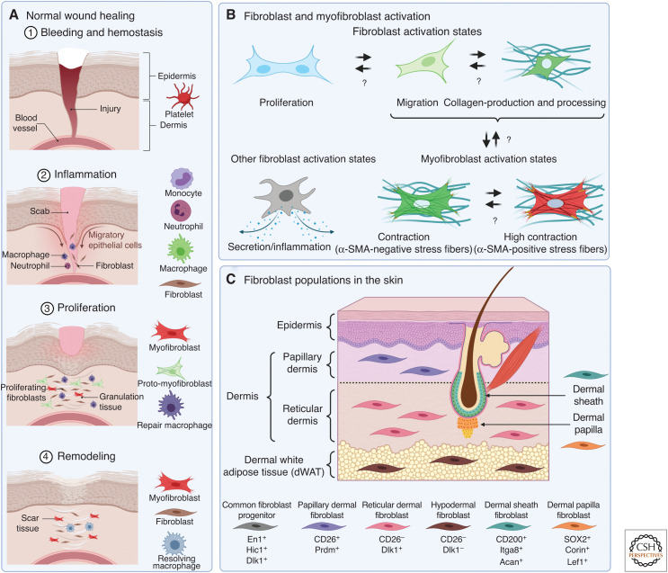
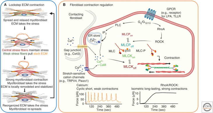
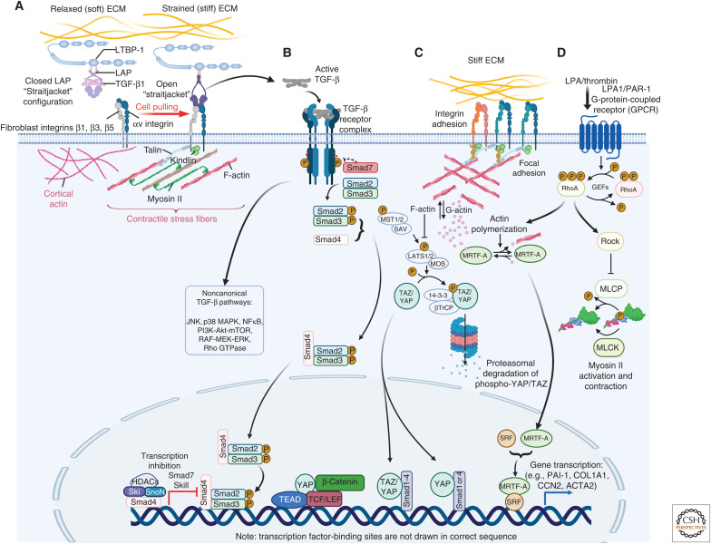
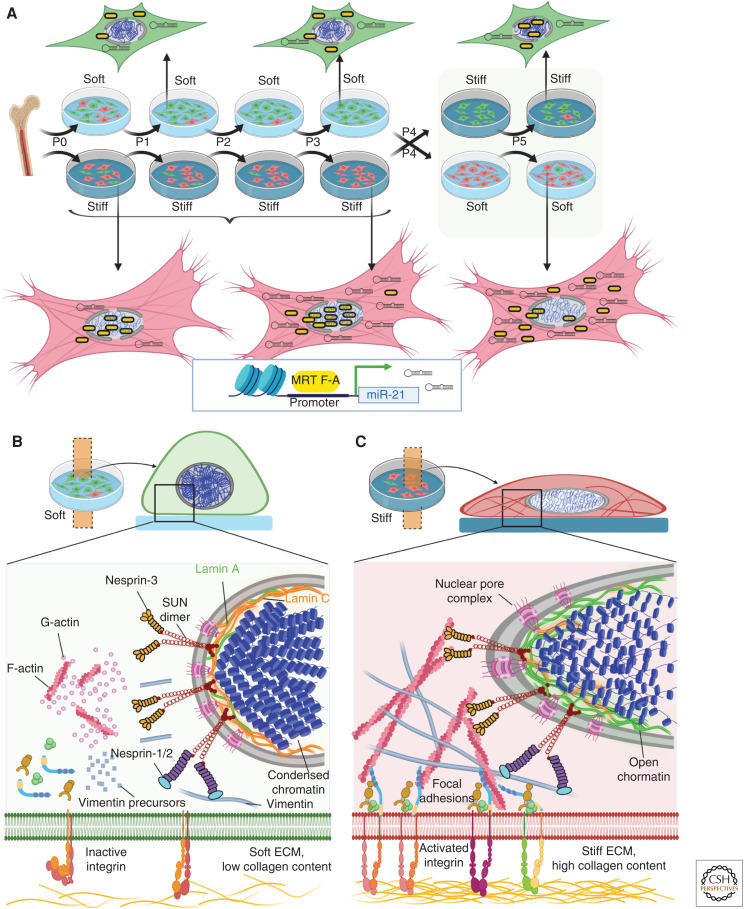
Myofibroblasts are the construction workers of wound healing and repair damaged tissues by producing and organizing collagen/extracellular matrix (ECM) into scar tissue. Scar tissue effectively and quickly restores the mechanical integrity of lost tissue architecture but comes at the price of lost tissue functionality. Fibrotic diseases caused by excessive or persistent myofibroblast activity can lead to organ failure. This review defines myofibroblast terminology, phenotypic characteristics, and functions. We will focus on the central role of the cell, ECM, and tissue mechanics in regulating tissue repair by controlling myofibroblast action. Additionally, we will discuss how therapies based on mechanical intervention potentially ameliorate wound healing outcomes. Although myofibroblast physiology and pathology affect all organs, we will emphasize cutaneous wound healing and hypertrophic scarring as paradigms for normal tissue repair versus fibrosis. A central message of this review is that myofibroblasts can be activated from multiple cell sources, varying with local environment and type of injury, to either restore tissue integrity and organ function or create an inappropriate mechanical environment.


Figures

Figure 1.
Figure 1.
Fibroblasts and myofibroblasts in the skin. (A) Positioning of different myofibroblast activation states in the time line and cellular context of normal skin wound healing. (B) Fibroblast activation is a loose term that can entail enhanced proliferation, migration, collagen production, or acquisition of a secretory and/or inflammatory phenotype. Such pre-activated fibroblasts can proceed to undergo myofibroblast activation, which is characterized by the formation of contractile stress fibers that can contain α-smooth muscle actin (α-SMA). There is documented reversibility and plasticity between the different activation states and phenotypes in vitro and in animal models of fibrosis and healing, indicated by bidirectional arrows. Note that not all dermal fibroblast precursors will be able to acquire all activation states. (C) Schematic representation different fibroblast populations that reside in dermal and subcutaneous tissues and their molecular markers. Created with BioRender.com.
Figure 2.
Figure 2.
Modes and regulation of fibroblast contraction. (A) In vitro studies support a biphasic cyclic lockstep mechanism of myofibroblast contraction that produces in collagen contractures over several days during normal wound healing to years in scar tissue. In stressed extracellular matrix (ECM) environments, µN-strong and long-lasting (hours) isometric contractions create slack in collagen fibrils, which thereby become accessible to local remodeling by fast (minutes), short-ranged, and pN-weak contractile events. Proteolytic processing of relaxed collagen fibers allows formation of shorter neo-ECM that takes over the mechanical load and allows myofibroblast re-spreading. (B) Strong isometric contraction is produced through RhoA/Rho-associated coiled-coil protein kinase (ROCK) activation, following binding of G-protein-coupled receptor (GPCR) ligands like lysophosphatic acid (LPA) to the LPA1 receptor or the thrombin cleavage product TLLR to the plasminogen-activated receptor (PAR)-1. RhoA-activated ROCK phosphorylates and thereby inhibits myosin light chain (MLC) phosphatase (MLCP), which mediates maintained actomyosin stress fiber contraction. Alternatively, short periodic contractions are regulated by transient increases in cytosolic Ca2+ through binding of calmodulin (CaM), which activates the MLC kinase (MLCK). High levels of cytosolic Ca2+ are achieved by calcium release from the endoplasmic reticulum following inositol trisphosphate (IP3) generation by phospholipase C (PLC) downstream of GPCR signaling. Other Ca2+ sources are adjacent cells connecting through gap junctions or Ca2+ entry from the extracellular milieu through stress-sensitive plasma membrane channels like transient receptor potential cation channel subfamily V member 4 (TRPV4) or piezo1. Created with BioRender.com.
Figure 3.
Figure 3.
Mechanical regulation of myofibroblast activation. (A) Association of TGF-β1 with its latency-associated peptide (LAP) creates a small latent complex (SLC) that is stored in the extracellular matrix (ECM) by binding to the latent TGF-β1-binding protein 1 (LTBP-1). A contractile cytoskeleton and mechanical resistance in the ECM shifts integrins αvβ1, and possibly αvβ3 and αvβ5 into the active conformation that enables pulling on RGD-binding sites present in LAP. If stiff and/or strained ECM resists the SLC straitjacket pully open to release active TGF-β1. (B) Active TGF-β1 binds to the TGF-β receptor complex to promote canonical Smad signaling or other signaling pathways, for example c-Jun amino-terminal kinase (JNK), p38, extracellular signal-regulated kinases (ERKs), mitogen-activated protein kinase (MAPK), and Rho-RhoA/Rho-associated coiled-coil protein kinase (ROCK). Biochemical and biomechanical myofibroblast signaling pathways converge at multiple intersection points. (C) RhoA-mediated ROCK activation contributes to the polymerization of G- into F-actin following integrin-mediated mechanosignaling, or (D) G-protein-coupled receptor (GPCR) ligand binding. Actin polymerization liberates myocardin-related transcription factor A (MRTF-A) from G-actin to translocate into the nucleus. Together with serum response factor (SRF), MRTF-A drives the transcription of profibrotic gene products, such as CCN2 and ACTA2 (α-SMA). ROCK further regulates gene expression through the transcriptional coactivators Yes-associated protein (YAP) and transcriptional coactivator with PDZ-binding motif (TAZ). Translocation of YAP and TAZ and association with transcription factors such TEA domain family member (TEAD) results in the transcription of genes that promote cell proliferation and fibrogenesis like CCN2 and miR-21. Created with BioRender.com.
Figure 4.
Figure 4.
Nuclear mechanics and myofibroblast mechanical memory. (A) Integrins in the cell membrane act as mechanosensors and are those in their inactive forms if not bound to extracellular ligands and in low affinity confirmations under low intracellular stress or soft extracellular matrix (ECM). In such low stress conditions, both actin microfilament and vimentin intermediate filament polymerization is low and connections with the nuclear lamina are weak. Low lamin A:C ratios in the inner nuclear membrane and condensed chromatin characterize fibroblasts grown in soft environments. With high intracellular stress and binding to stiff ECM with high collagen content, integrins shift to their active confirmation and cluster into multicomponent focal adhesion structures. (B) Mechanotransduction pathways and stress originating at cell–ECM adhesions drive polymerization of G- to F-actin and formation of vimentin intermediate filaments that establish a direct connection between ECM adhesions and the linker of the nucleoskeleton and cytoskeleton (LINC) complex in the nuclear outer membrane. The LINC complex contains nuclear envelope spectrin repeat proteins (nesprins), and Sad1p and UNC-84 homology (SUN) proteins that span the nuclear envelope. Nesprin-3 connect SUN proteins to F-actin, whereas nesprins-1 and -2 connect to intermediate filaments. On the inner nuclear membrane, SUN dimers interact with lamin A attached to chromatin, which is thus forced to open in high stress conditions. (C) Imprinting of in vitro mechanical memory has been demonstrated for mesenchymal stromal cells (MSCs) directly isolated from bone marrow onto soft (5 kPa elastic modulus) and stiff (100 kPa) culture substrates. By continuously passaging on stiff or soft substrates (P = one passage of 7 d), MSCs are mechanically primed. On stiff substrates, the mechanosensitive myocardin-related transcription factor A (MRTF-A) localizes to the nucleus and drives continued transcription of the profibrotic micro-RNA miR-21; MRTF-A remains cytosolic on soft substrates. After the switch from stiff to soft substrates, MRTF-A acutely shifts to the cytosol, whereas miR-21 levels remain high due to low turnover rates and continue to promote myofibroblast activation for at least 2 more weeks as one keeper of mechanical memory. Created with BioRender.com.

Similar articles

Cited by

References

    1. Aarabi S, Bhatt KA, Shi Y, Paterno J, Chang EI, Loh SA, Holmes JW, Longaker MT, Yee H, Gurtner GC. 2007. Mechanical load initiates hypertrophic scar formation through decreased cellular apoptosis. FASEB J 21: 3250–3261. 10.1096/fj.07-8218com - DOI - PubMed
    1. Abbasi S, Sinha S, Labit E, Rosin NL, Yoon G, Rahmani W, Jaffer A, Sharma N, Hagner A, Shah P, et al. 2020. Distinct regulatory programs control the latent regenerative potential of dermal fibroblasts during wound healing. Cell Stem Cell 27: 396–412.e396. 10.1016/j.stem.2020.07.008 - DOI - PubMed
    1. Abercrombie M, James DW, Newcombe JF. 1960. Wound contraction in rabbit skin, studied by splinting the wound margins. J Anat 94: 170–182. - PMC - PubMed
    1. Achterberg VF, Buscemi L, Diekmann H, Smith-Clerc J, Schwengler H, Meister JJ, Wenck H, Gallinat S, Hinz B. 2014. The nano-scale mechanical properties of the extracellular matrix regulate dermal fibroblast function. J Invest Dermatol 134: 1862–1872. 10.1038/jid.2014.90 - DOI - PubMed
    1. Adapala RK, Katari V, Teegala LR, Thodeti S, Paruchuri S, Thodeti CK. 2021. TRPV4 mechanotransduction in fibrosis. Cells 10: 3053. 10.3390/cells10113053 - DOI - PMC - PubMed

Publication types

LinkOut - more resources