Review
. 2023 Feb;25(1):36-45.
doi: 10.1007/s11307-022-01772-8. Epub 2022 Sep 19.

The Evolution of Fluorescence-Guided Surgery

Affiliations
Review

The Evolution of Fluorescence-Guided Surgery

Stan Van Keulen et al. Mol Imaging Biol. 2023 Feb.

Abstract

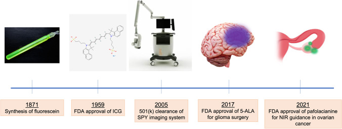
There has been continual development of fluorescent agents, imaging systems, and their applications over the past several decades. With the recent FDA approvals of 5-aminolevulinic acid, hexaminolevulinate, and pafolacianine, much of the potential that fluorescence offers for image-guided oncologic surgery is now being actualized. In this article, we review the evolution of fluorescence-guided surgery, highlight the milestones which have contributed to successful clinical translation, and examine the future of targeted fluorescence imaging.

Keywords: Fluorescence-guided surgery; Molecular imaging; Oncology.


Conflict of interest statement

E.L.R. has equipment loans from Stryker and consults for Rakuten Medical. All other authors declare that they have no conflict of interest.

Figures

Fig. 1
Fig. 1
Milestones in the evolution of fluorescence-guided surgery. Fluorescein was initially discovered in the middle of the nineteenth century, but ICG became the first FDA-approved fluorescent dye. Subsequent clearance of the Novodaq SPY imaging system paved the way for device development, and a growing clinical experience has provided the foundation for FDA approval of fluorescent agents for intraoperative use.
Fig. 2
Fig. 2
ICG angiography in microvascular reconstructive surgery. A Identification of a perforator is crucial during an anterolateral thigh free flap harvest but can be challenging with reflected light visualization only. B The perforator can be readily identified (arrow) through its course in muscle by NIR imaging. ICG angiography is also useful in assessment of flow through a microvascular anastomosis. C By reflected light imaging, venous flow (arrow) is difficult to ascertain. D By ICG angiography and SPY NIR imaging, the artery is observed to have adequate flow (arrowhead), but the venous flow (arrow) is insufficient (D). E Restoration of venous flow subsequently confirmed by ICG imaging (arrow).
Fig. 3
Fig. 3
Assessment of tissue perfusion with SPY NIR imaging using intravenous ICG infusion. A The right lingual artery was ligated during an oncologic head and neck surgery. Vascularity to the right hemitongue (arrows) appears normal on reflected light images. B However, a complete absence of vascular flow is apparent on ICG angiography (B).
Fig. 4
Fig. 4
Sentinel lymph node identification using ICG. A A total of 5 mg of ICG was injected submucosally around a left tongue squamous cell carcinoma (arrowheads). B The neck was subsequently imaged using a handheld SPY NIR imaging system, and a submental lymph node (arrowhead) was identified by fluorescence.
Fig. 5
Fig. 5
Fluorescence-guided surgery workflow. First, the fluorescence tracer is venously infused preoperatively. During surgery, in vivo open-field camera systems are used to visualize tumor tissue, and to assess adjacent tissue and the wound bed. After the tumor resection, the tumor specimen (i.e., the tumor with a cuff of healthy surrounding tissue) is imaged within a closed-field ex vivo fashion. Thereafter, during histological assessment, the fluorescence is traced within the tumor specimen to ascertain if fluorescence areas correspond with tumor areas.
Fig. 6
Fig. 6
Activatable fluorescence probes. A Enzyme-activable probes allow for fluorescence signal to emit only in the presence of specific enzymatic activity. B An alternative strategy allows for fluorescence activation after incorporation of the agent by surface molecule binding and lysosome phagocytosis by the targeted cancer cell.
Fig. 7
Fig. 7
pH induced fluorescence agent activation. The agent is in quenched state within a micelle in a pH neutral environment. As the micelle reaches the acidic tumor environment, it dissolves and the agent starts to emit its photons when excitation takes place.

References

    1. Orosco RK, Tapia VJ, Califano JA, Clary B, Cohen EEW, Kane C, et al. Positive surgical margins in the 10 most common solid cancers. Sci Rep. 2018;8(1):5686. doi: 10.1038/s41598-018-23403-5. - DOI - PMC - PubMed
    1. Woolgar JA, Triantafyllou A. A histopathological appraisal of surgical margins in oral and oropharyngeal cancer resection specimens. Oral Oncol. 2005;41(10):1034–1043. doi: 10.1016/j.oraloncology.2005.06.008. - DOI - PubMed
    1. Atkins J, Al Mushawah F, Appleton CM, Cyr AE, Gillanders WE, Aft RL, et al. Positive margin rates following breast-conserving surgery for stage I-III breast cancer: palpable versus nonpalpable tumors. J Surg Res. 2012;177(1):109–115. doi: 10.1016/j.jss.2012.03.045. - DOI - PMC - PubMed
    1. Iczkowski KA, Lucia MS. Frequency of positive surgical margin at prostatectomy and its effect on patient outcome. Prostate Cancer. 2011;2011:673021. doi: 10.1155/2011/673021. - DOI - PMC - PubMed
    1. Hadjipanayis CG, Stummer W. 5-ALA and FDA approval for glioma surgery. J Neurooncol. 2019;141(3):479–486. doi: 10.1007/s11060-019-03098-y. - DOI - PMC - PubMed

LinkOut - more resources